Nuffield Department of Medicine, University of Oxford, The Peter Medawar Building for Pathogen Research, South Parks Road, Oxford OX1 3SY, UK.
BMC Med Genomics. 2010 Oct 13;3:46. doi: 10.1186/1755-8794-3-46.
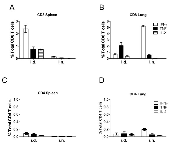
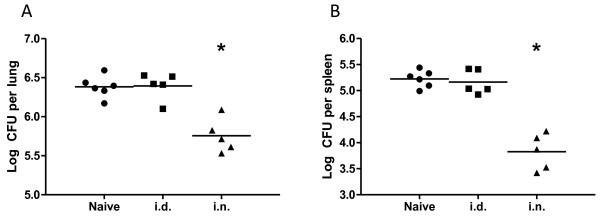
Immunization of BALB/c mice with a recombinant adenovirus expressing Mycobacterium tuberculosis (M. tuberculosis) antigen 85A (Ad85A) protects against aerosol challenge with M. tuberculosis only when it is administered intra-nasally (i.n.). Immunization with Ad85A induces a lung-resident population of activated CD8 T cells that is antigen dependent, highly activated and mediates protection by early inhibition of M. tuberculosis growth. In order to determine why the i.n. route is so effective compared to parenteral immunization, we used microarray analysis to compare gene expression profiles of pulmonary and splenic CD8 T cells after i.n. or intra-dermal (i.d.) immunization.
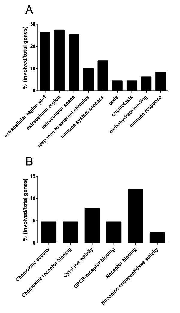
Total RNA from CD8 T cells was isolated from lungs or spleens of mice immunized with Ad85A by the i.n. or i.d. route. The gene profiles generated from each condition were compared. Statistically significant (p ≤ 0.05) differentially expressed genes were analyzed to determine if they mapped to particular molecular functions, biological processes or pathways using Gene Ontology and Panther DB mapping tools.
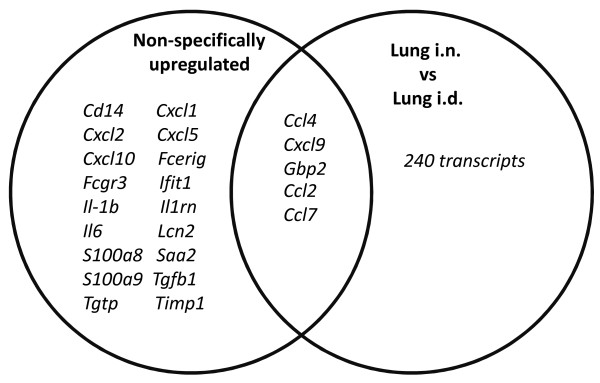
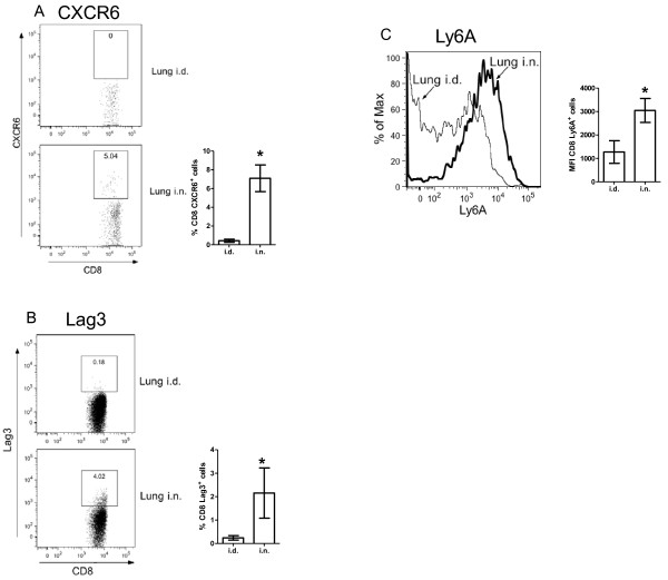
CD8 T cells from lungs of i.n. immunized mice expressed a large number of chemokines chemotactic for resting and activated T cells as well as activation and survival genes. Lung lymphocytes from i.n. immunized mice also express the chemokine receptor gene Cxcr6, which is thought to aid long-term retention of antigen-responding T cells in the lungs. Expression of CXCR6 on CD8 T cells was confirmed by flow cytometry.
Our microarray analysis represents the first ex vivo study comparing gene expression profiles of CD8 T cells isolated from distinct sites after immunization with an adenoviral vector by different routes. It confirms earlier phenotypic data indicating that lung i.n. cells are more activated than lung i.d. CD8 T cells. The sustained expression of chemokines and activation genes enables CD8 T cells to remain in the lungs for extended periods after i.n. immunization. This may account for the early inhibition of M. tuberculosis growth observed in Ad85A i.n. immunized mice and explain the effectiveness of i.n. compared to parenteral immunization with this viral vector.
用表达结核分枝杆菌(M. tuberculosis)抗原 85A(Ad85A)的重组腺病毒免疫 BALB/c 小鼠,仅当经鼻内(i.n.)途径给予时,可预防雾化挑战中的结核分枝杆菌。用 Ad85A 免疫诱导肺驻留的激活 CD8 T 细胞群体,该群体依赖抗原,高度激活,并通过早期抑制结核分枝杆菌生长来介导保护。为了确定与肠外免疫相比,为什么 i.n. 途径如此有效,我们使用微阵列分析比较了经 i.n.或皮内(i.d.)免疫后肺和脾 CD8 T 细胞的基因表达谱。
用 Ad85A 经 i.n.或 i.d.途径免疫的小鼠的 CD8 T 细胞从肺或脾中分离总 RNA。比较每个条件下生成的基因谱。使用基因本体论和 Panther DB 映射工具分析统计上显著(p ≤ 0.05)差异表达的基因,以确定它们是否映射到特定的分子功能、生物学过程或途径。
经 i.n.免疫小鼠肺 CD8 T 细胞表达了大量趋化因子,可趋化静止和激活的 T 细胞以及激活和存活基因。经 i.n.免疫小鼠的肺淋巴细胞也表达趋化因子受体基因 Cxcr6,这被认为有助于抗原反应性 T 细胞在肺部的长期保留。通过流式细胞术证实了 CXCR6 在 CD8 T 细胞上的表达。
我们的微阵列分析代表了首次通过不同途径用腺病毒载体免疫后比较从不同部位分离的 CD8 T 细胞的基因表达谱的体外研究。它证实了早期表型数据表明,肺内 CD8 T 细胞比肺内 i.d. CD8 T 细胞更活跃。趋化因子和激活基因的持续表达使 CD8 T 细胞在经 i.n.免疫后能在肺部持续存在较长时间。这可能解释了在 Ad85A i.n.免疫的小鼠中观察到的结核分枝杆菌生长的早期抑制,并解释了与这种病毒载体的肠外免疫相比,i.n. 免疫的有效性。