Institute of Condensed Matter and Nanosciences--Bio & Soft Matter, Université catholique de Louvain, Croix du Sud 2/18, Louvain-la-Neuve B-1348, Belgium.
Nat Commun. 2010 Jun 15;1(3):27. doi: 10.1038/ncomms1027.
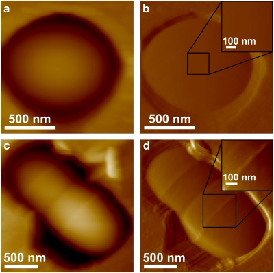
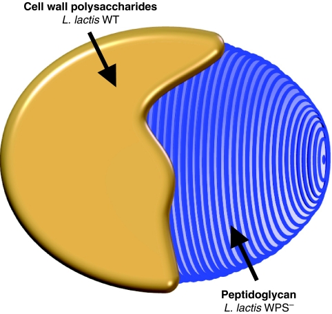
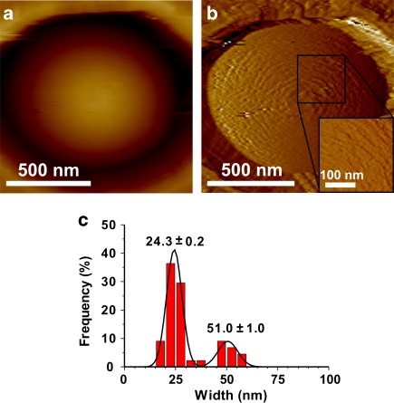
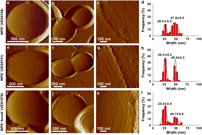
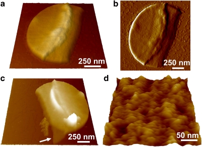
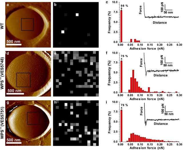
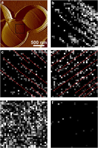
The spatial organization of peptidoglycan, the major constituent of bacterial cell-walls, is an important, yet still unsolved issue in microbiology. In this paper, we show that the combined use of atomic force microscopy and cell wall mutants is a powerful platform for probing the nanoscale architecture of cell wall peptidoglycan in living Gram-positive bacteria. Using topographic imaging, we found that Lactococcus lactis wild-type cells display a smooth, featureless surface morphology, whereas mutant strains lacking cell wall exopolysaccharides feature 25-nm-wide periodic bands running parallel to the short axis of the cell. In addition, we used single-molecule recognition imaging to show that parallel bands are made of peptidoglycan. Our data, obtained for the first time on living ovococci, argue for an architectural feature of the cell wall in the plane perpendicular to the long axis of the cell. The non-invasive live cell experiments presented here open new avenues for understanding the architecture and assembly of peptidoglycan in Gram-positive bacteria.
肽聚糖是细菌细胞壁的主要成分,其空间组织是微生物学中一个重要但尚未解决的问题。在本文中,我们表明,原子力显微镜和细胞壁突变体的联合使用是探测活革兰氏阳性细菌细胞壁肽聚糖纳米结构的强大平台。通过形貌成像,我们发现乳球菌属野生型细胞呈现出光滑、无特征的表面形态,而缺乏细胞壁胞外多糖的突变株则具有 25nm 宽的、与细胞短轴平行的周期性带。此外,我们还利用单分子识别成像表明平行带由肽聚糖组成。我们的数据首次在活卵形球菌上获得,证明了细胞壁在垂直于细胞长轴的平面上的结构特征。本文提出的非侵入性活细胞实验为理解革兰氏阳性细菌中肽聚糖的结构和组装开辟了新途径。