Molecular Physiology and Biophysics Section, Porter Neuroscience Research Center, National Institute of Neurological Disorders and Stroke, National Institutes of Health, Bethesda, Maryland 20892, USA.
Nat Commun. 2010 Jul 27;1(4):44. doi: 10.1038/ncomms1048.
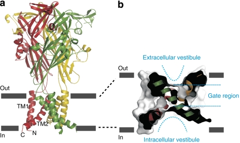
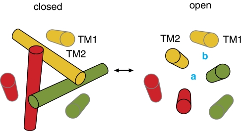
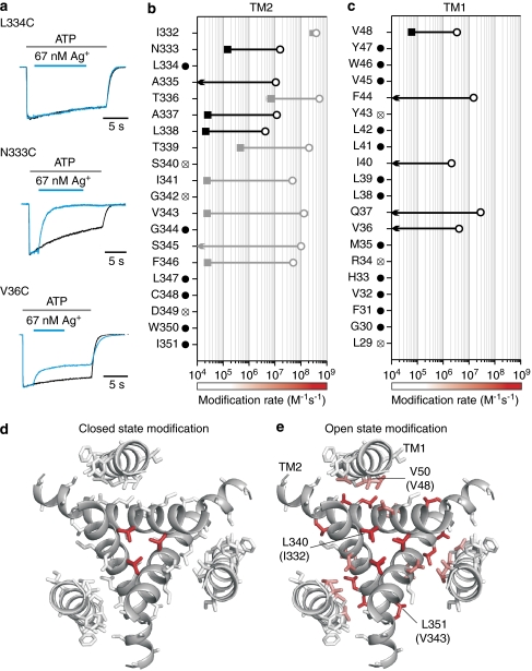
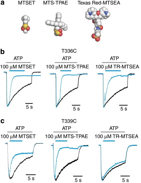
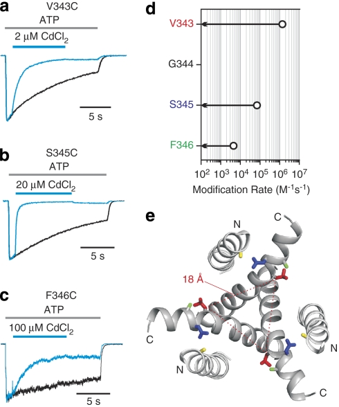
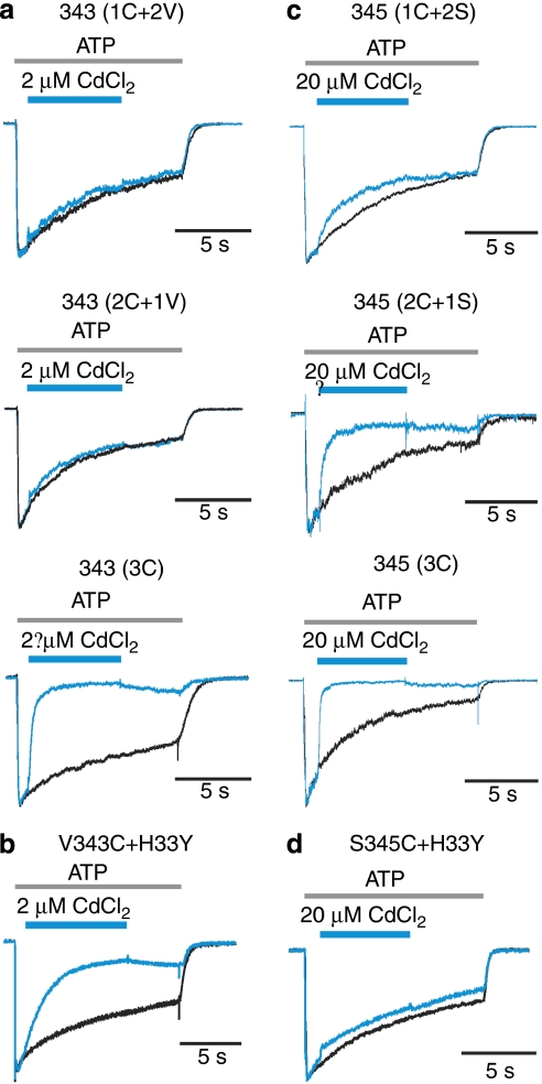
The opening of ion channels in response to ligand binding, voltage or membrane stretch underlies electrical and chemical signalling throughout biology. Two structural classes of pore-opening mechanisms have been established, including bending of pore-lining helices in the case of tetrameric cation channels, or tilting of such helices in mechanosensitive channels. In this paper, we explore how the structure of the pore changes during opening in P2X receptors by measuring the modification of introduced cysteine residues in transmembrane helices by thiol-reactive reagents, and by engineering metal bridges. Our results are consistent with the X-ray structure of the closed state, and demonstrate that expansion of the gate region in the external pore is accompanied by a significant narrowing of the inner pore, indicating that pore-forming helices straighten on ATP binding to open the channel. This unique pore-opening mechanism has fundamental implications for the role of subunit interfaces in the gating mechanism of P2X receptors and points to a role of the internal pore in ion permeation.
配体结合、电压或膜拉伸引起的离子通道开放是整个生物学中电信号和化学信号的基础。已经确定了两种结构类别的孔开启机制,包括四聚体阳离子通道中孔衬螺旋的弯曲,或机械敏感通道中这种螺旋的倾斜。在本文中,我们通过测量跨膜螺旋中引入的半胱氨酸残基被巯基反应试剂修饰的情况以及通过工程金属桥的方法,来研究 P2X 受体在开放过程中孔结构如何发生变化。我们的结果与关闭状态的 X 射线结构一致,并表明外孔门区的扩张伴随着内孔的明显变窄,表明在 ATP 结合打开通道时,形成孔的螺旋伸直。这种独特的孔开启机制对 P2X 受体门控机制中亚基界面的作用具有根本意义,并指出了内部孔在离子渗透中的作用。