University of Giessen Lung Centre, Giessen, Germany.
Respir Res. 2010 Oct 27;11(1):146. doi: 10.1186/1465-9921-11-146.
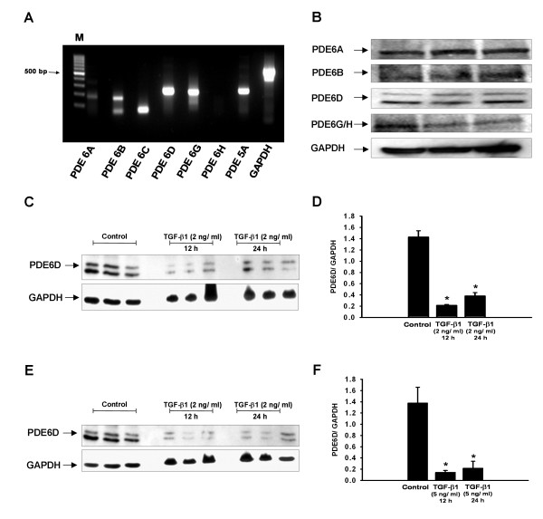
Idiopathic Pulmonary Fibrosis (IPF) is an unresolved clinical issue. Phosphodiesterases (PDEs) are known therapeutic targets for various proliferative lung diseases. Lung PDE6 expression and function has received little or no attention. The present study aimed to characterize (i) PDE6 subunits expression in human lung, (ii) PDE6 subunits expression and alteration in IPF and (iii) functionality of the specific PDE6D subunit in alveolar epithelial cells (AECs).
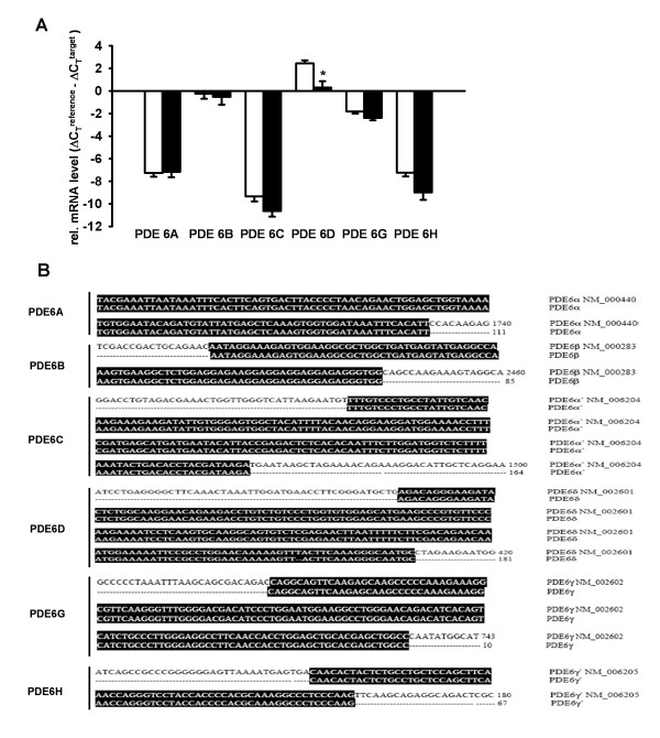
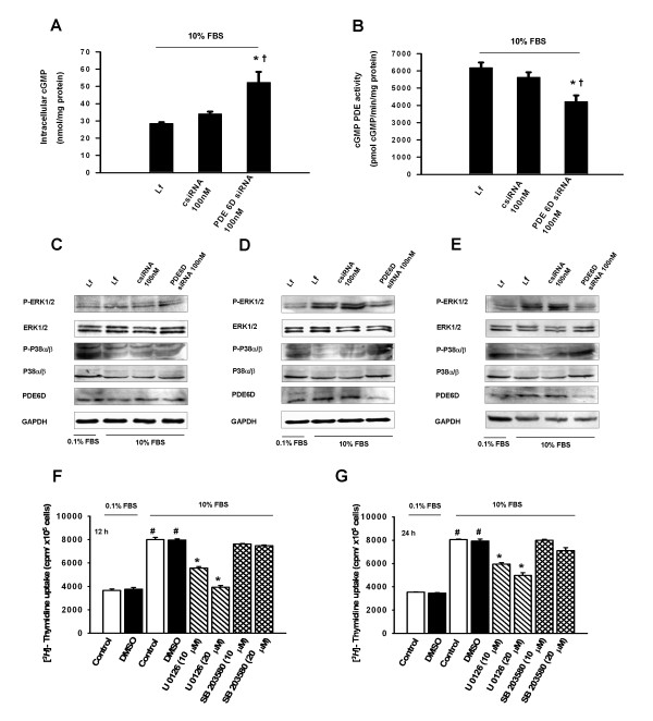
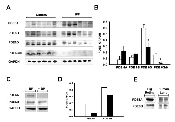
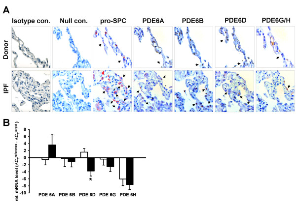
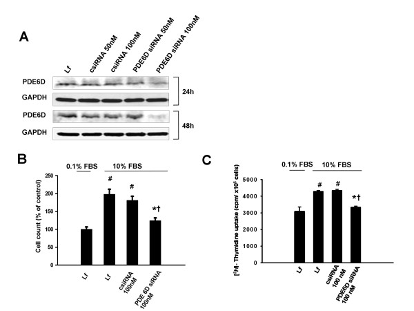
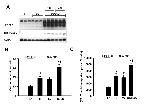
METHODOLOGY/PRINCIPAL FINDINGS: PDE6 subunits expression in transplant donor (n = 6) and IPF (n = 6) lungs was demonstrated by real-time quantitative (q)RT-PCR and immunoblotting analysis. PDE6D mRNA and protein levels and PDE6G/H protein levels were significantly down-regulated in the IPF lungs. Immunohistochemical analysis showed alveolar epithelial localization of the PDE6 subunits. This was confirmed by qRT-PCR from human primary alveolar type (AT)II cells, demonstrating the down-regulation pattern of PDE6D in IPF-derived ATII cells. In vitro, PDE6D protein depletion was provoked by transforming growth factor (TGF)-β1 in A549 AECs. PDE6D siRNA-mediated knockdown and an ectopic expression of PDE6D modified the proliferation rate of A549 AECs. These effects were mediated by increased intracellular cGMP levels and decreased ERK phosphorylation.
CONCLUSIONS/SIGNIFICANCE: Collectively, we report previously unrecognized PDE6 expression in human lungs, significant alterations of the PDE6D and PDE6G/H subunits in IPF lungs and characterize the functional role of PDE6D in AEC proliferation.
特发性肺纤维化(IPF)是一个未解决的临床问题。磷酸二酯酶(PDEs)是各种增殖性肺疾病的已知治疗靶点。肺 PDE6 表达和功能尚未得到充分关注。本研究旨在(i)描述人肺中 PDE6 亚基的表达,(ii)描述 IPF 中 PDE6 亚基的表达和改变,以及(iii)肺泡上皮细胞(AECs)中特定 PDE6D 亚基的功能。
方法/主要发现:通过实时定量(q)RT-PCR 和免疫印迹分析,证明了移植供体(n = 6)和 IPF (n = 6)肺中 PDE6 亚基的表达。在 IPF 肺中,PDE6D mRNA 和蛋白水平以及 PDE6G/H 蛋白水平显著下调。免疫组织化学分析显示 PDE6 亚基在肺泡上皮细胞中的定位。这通过来自人原代肺泡型(AT)II 细胞的 qRT-PCR 得到证实,证明了 IPF 衍生的 ATII 细胞中 PDE6D 的下调模式。在体外,转化生长因子(TGF)-β1 可引起 A549 AEC 中 PDE6D 蛋白耗竭。PDE6D siRNA 介导的敲低和 PDE6D 的异位表达改变了 A549 AEC 的增殖率。这些作用是通过增加细胞内 cGMP 水平和降低 ERK 磷酸化来介导的。
结论/意义:总之,我们报告了人肺中以前未被识别的 PDE6 表达、IPF 肺中 PDE6D 和 PDE6G/H 亚基的显著改变,并描述了 PDE6D 在 AEC 增殖中的功能作用。