Department of Traditional and Western Medical Hepatology, Third Hospital of Hebei Medical University, Shijiazhuang, PR China.
Lipids Health Dis. 2010 Oct 28;9:124. doi: 10.1186/1476-511X-9-124.
Heme oxygenase-1 (HO-1), the rate-limiting enzyme in heme catabolism, has been reported to have potential antioxidant properties. However, the role of HO-1 on hepatocyte apoptosis remains unclear. We aim to elucidate the effects of HO-1 on oxidative stress related hepatocellular apoptosis in nutritional steatohepatitis in mice.
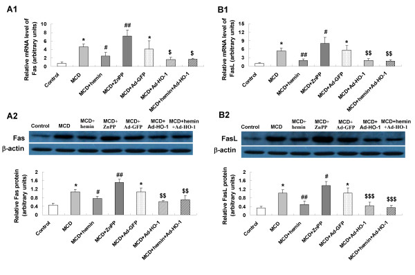
C57BL/6J mice were fed with methionine-choline deficient (MCD) diet for four weeks to induce hepatic steatohepatitis. HO-1 chemical inducer (hemin), HO-1 chemical inhibitor zinc protoporphyrin IX (ZnPP-IX) and/or adenovirus carrying HO-1 gene (Ad-HO-1) were administered to mice, respectively. Hepatocyte apoptosis was evaluated by terminal deoxynucleotidyl transferase dUTP nick-end labeling (TUNEL) assay, the mRNA and protein expression of apoptosis related genes were assayed by quantitative real-time PCR and Western blot.
Hepatocyte signs of oxidative related apoptotic injury were presented in mice fed with MCD diet for 4 weeks. Induction of HO-1 by hemin or Ad-HO-1 significantly attenuated the severity of liver histology, which was associated with decreased hepatic lipid peroxidation content, reduced number of apoptotic cells by TUNEL staining, down-regulated expression of pro-apoptosis related genes including Fas/FasL, Bax, caspase-3 and caspase-9, reduced expression of cytochrome p4502E1 (CYP2E1), inhibited cytochrome c (Cyt-c) release, and up-regulated expression of anti-apoptosis gene Bcl-2. Whereas, inhibition of HO-1 by ZnPP-IX caused oxidative stress related hepatic injury, which concomitant with increased number of TUNEL positive cells and up-regulated expression of pro-apoptosis related genes.
The present study provided evidences for the protective role of HO-1 in preventing nutritional steatohepatitis through suppressing hepatocyte apoptosis in mice.
血红素加氧酶-1(HO-1)是血红素分解代谢的限速酶,具有潜在的抗氧化特性。然而,HO-1 对肝细胞凋亡的作用尚不清楚。本研究旨在阐明 HO-1 在营养性脂肪性肝炎小鼠氧化应激相关肝细胞凋亡中的作用。
C57BL/6J 小鼠给予蛋氨酸-胆碱缺乏(MCD)饮食 4 周诱导肝脂肪性肝炎。分别给予 HO-1 化学诱导剂(血红素)、HO-1 化学抑制剂锌原卟啉 IX(ZnPP-IX)和/或携带 HO-1 基因的腺病毒(Ad-HO-1)。通过末端脱氧核苷酸转移酶 dUTP 缺口末端标记(TUNEL)检测法评估肝细胞凋亡,通过实时定量 PCR 和 Western blot 检测凋亡相关基因的 mRNA 和蛋白表达。
给予 MCD 饮食 4 周的小鼠出现肝细胞氧化相关凋亡损伤迹象。血红素或 Ad-HO-1 诱导 HO-1 可显著减轻肝组织学严重程度,同时降低肝脂质过氧化含量,减少 TUNEL 染色的凋亡细胞数,下调 Fas/FasL、Bax、caspase-3 和 caspase-9 等促凋亡相关基因的表达,下调细胞色素 P4502E1(CYP2E1)表达,抑制细胞色素 c(Cyt-c)释放,上调抗凋亡基因 Bcl-2 的表达。相反,ZnPP-IX 抑制 HO-1 导致氧化应激相关肝损伤,同时增加 TUNEL 阳性细胞数,并上调促凋亡相关基因的表达。
本研究为 HO-1 通过抑制小鼠肝细胞凋亡在预防营养性脂肪性肝炎中的保护作用提供了证据。