Division of Cardiovascular Medicine, University of Florida College of Medicine, Gainesville, Florida, USA.
J Interv Cardiol. 2010 Dec;23(6):511-9. doi: 10.1111/j.1540-8183.2010.00598.x. Epub 2010 Oct 4.
Using intravascular ultrasound (IVUS), we sought to characterize coronary morphology in women with chest pain without major epicardial obstructive coronary artery disease (CAD). We have previously observed an unexpectedly high rate of adverse outcomes among women with chest pain and normal or insignificant obstructive CAD. Information about the presence and characteristics of coronary atherosclerosis in these women could provide insight into the mechanisms related to increased risk, as well as improved diagnosis, prevention, and treatment.
Women (n = 100) with suspected ischemia without obstructive CAD (>50% stenosis) underwent IVUS of a left coronary segment with measurements by a core lab masked to clinical and angiographic findings.
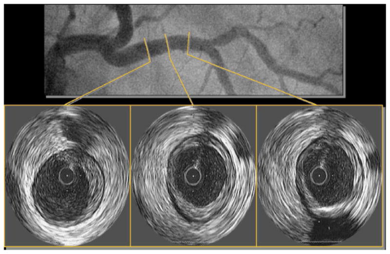
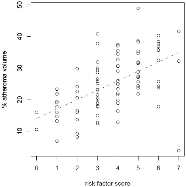
Angiograhic core lab analysis found 69.6% of patients had no (≤20%) and 30.4% had minimal (20-<50%) CAD. IVUS segmental images were interpretable by the core lab in 92 women, with 19 (21%) having no atherosclerosis (intimal-medial thickness <0.5 mm). In the remaining 73 women (79%), percent atheroma volume was 27 ± 8% and mean maximum plaque thickness was 0.53 ± 0.22 mm. Thirty-eight women with atherosclerosis (53%) had ≥30% of interrogated vessel involved. The average vessel involvement was 40%, and the maximum plaque thickness was 1.27 mm. The number of risk factors strongly correlated with percent atheroma volume (r = 0.53, P < 0.0001) and percent vessel involvement (r = 0.51, P < 0.0001), with the strongest independent predictor of both being age. Remodeling was assessed in 59/73 women (81%), and 73% had evidence of positive remodeling.
In symptomatic women without significant luminal obstructive CAD, we observed a high prevalence of atherosclerosis with positive remodeling and preserved lumen size. These findings may help explain increased risk and emphasize need for improved diagnostic and treatment options for women with concealed CAD.
通过血管内超声(IVUS),我们试图描述胸痛但无主要心外膜阻塞性冠状动脉疾病(CAD)的女性的冠状动脉形态。我们之前观察到,胸痛且 CAD 无阻塞或轻度阻塞的女性不良结局发生率出乎意料地高。这些女性的冠状动脉粥样硬化的存在和特征信息可能有助于深入了解与风险增加相关的机制,并改善诊断、预防和治疗。
100 名疑似缺血但无阻塞性 CAD(>50%狭窄)的女性接受了左冠状动脉节段的 IVUS 检查,其测量结果由一个核心实验室进行,核心实验室对临床和血管造影发现进行了盲法分析。
血管造影核心实验室分析发现,69.6%的患者无(≤20%)和 30.4%的患者有轻度(20-<50%)CAD。核心实验室可对 92 名女性的 IVUS 节段图像进行解释,其中 19 名(21%)无动脉粥样硬化(内膜-中层厚度<0.5 毫米)。在其余 73 名女性(79%)中,粥样斑块体积百分比为 27±8%,最大斑块厚度为 0.53±0.22 毫米。38 名动脉粥样硬化女性(53%)有≥30%的受检血管受累。平均血管受累程度为 40%,最大斑块厚度为 1.27 毫米。危险因素的数量与粥样斑块体积百分比(r=0.53,P<0.0001)和血管受累百分比(r=0.51,P<0.0001)呈强相关性,年龄是两者最强的独立预测因素。在 59/73 名女性(81%)中评估了重构,其中 73%有正性重构的证据。
在无明显管腔阻塞性 CAD 的有症状女性中,我们观察到动脉粥样硬化的高患病率,伴有正性重构和管腔大小保持不变。这些发现可能有助于解释风险增加,并强调需要为隐匿性 CAD 女性提供更好的诊断和治疗选择。