Institute of Cellular Biology and Pathology 'Nicolae Simionescu', Bucharest, Romania Institute for Neurophysiology, University of Cologne, Cologne, Germany.
J Cell Mol Med. 2011 Sep;15(9):1914-26. doi: 10.1111/j.1582-4934.2010.01197.x.
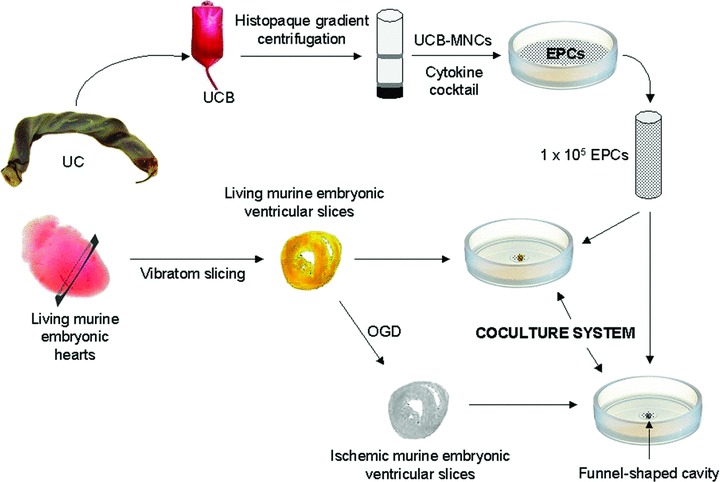
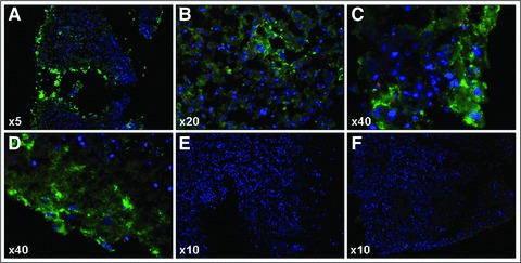
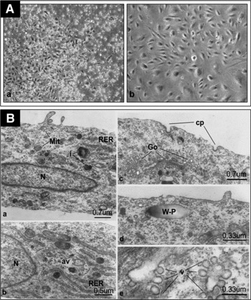
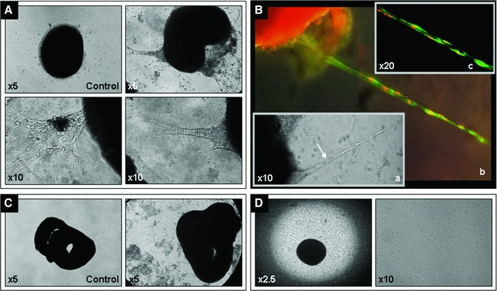
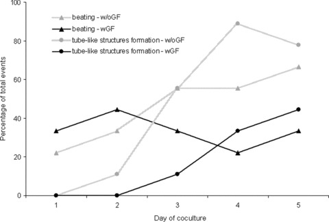
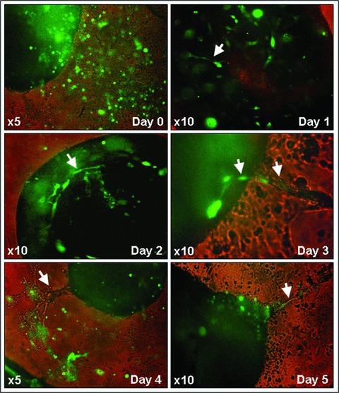
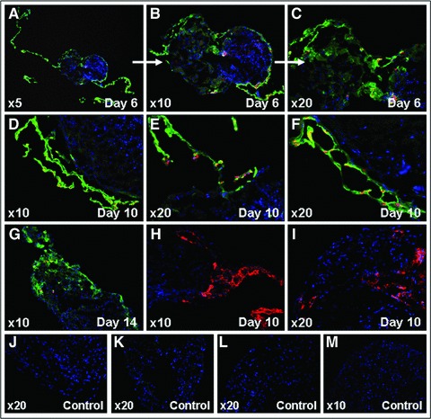
The umbilical cord blood derived endothelial progenitor cells (EPCs) contribute to vascular regeneration in experimental models of ischaemia. However, their ability to participate in cardiovascular tissue restoration has not been elucidated yet. We employed a novel coculture system to investigate whether human EPCs have the capacity to integrate into living and ischaemic cardiac tissue, and participate to neovascularization. EPCs were cocultured with either living or ischaemic murine embryonic ventricular slices, in the presence or absence of a pro-angiogenic growth factor cocktail consisting of VEGF, IGF-1, EGF and bFGF. Tracking of EPCs within the cocultures was performed by cell transfection with green fluorescent protein or by immunostaining performed with anti-human vWF, CD31, nuclei and mitochondria antibodies. EPCs generated vascular tube-like structures in direct contact with the living ventricular slices. Furthermore, the pro-angiogenic growth factor cocktail reduced significantly tubes formation. Coculture of EPCs with the living ventricular slices in a transwell system did not lead to vascular tube-like structures formation, demonstrating that the direct contact is necessary and that the soluble factors secreted by the living slices were not sufficient for their induction. No vascular tubes were formed when EPCs were cocultured with ischaemic ventricular slices, even in the presence of the pro-angiogenic cocktail. In conclusion, EPCs form vascular tube-like structures in contact with living cardiac tissue and the direct cell-to-cell interaction is a prerequisite for their induction. Understanding the cardiac niche and micro-environmental interactions that regulate EPCs integration and neovascularization are essential for applying these cells to cardiovascular regeneration.
脐带血衍生的内皮祖细胞 (EPCs) 有助于缺血实验模型中的血管再生。然而,它们参与心血管组织修复的能力尚未阐明。我们采用了一种新的共培养系统来研究人类 EPC 是否有能力整合到活体和缺血性心肌组织中,并参与血管新生。将 EPC 与活体或缺血性的鼠胚胎心室片共培养,共培养物中存在或不存在由 VEGF、IGF-1、EGF 和 bFGF 组成的促血管生成生长因子鸡尾酒。通过用绿色荧光蛋白转染细胞或用抗人 vWF、CD31、核和线粒体抗体进行免疫染色来追踪共培养物中的 EPC。EPC 与活体心室片直接接触时会生成血管管状结构。此外,促血管生成生长因子鸡尾酒显著减少了管腔形成。EPC 与活体心室片在 Transwell 系统中共培养不会导致血管管状结构形成,这表明直接接触是必需的,并且活体切片分泌的可溶性因子不足以诱导其形成。当 EPC 与缺血性心室片共培养时,即使存在促血管生成鸡尾酒,也不会形成血管管状结构。总之,EPC 与活体心肌组织接触时会形成血管管状结构,而直接的细胞间相互作用是诱导其形成的前提。了解调节 EPC 整合和血管新生的心脏龛位和微环境相互作用对于将这些细胞应用于心血管再生至关重要。