• 文献检索
  • 文档翻译
  • 深度研究
  • 学术资讯
  • Suppr Zotero 插件Zotero 插件
  • 邀请有礼
  • 套餐&价格
  • 历史记录
应用&插件
Suppr Zotero 插件Zotero 插件浏览器插件Mac 客户端Windows 客户端微信小程序
定价
高级版会员购买积分包购买API积分包
服务
文献检索文档翻译深度研究API 文档MCP 服务
关于我们
关于 Suppr公司介绍联系我们用户协议隐私条款
关注我们

Suppr 超能文献

核心技术专利:CN118964589B侵权必究
粤ICP备2023148730 号-1Suppr @ 2026

文献检索

告别复杂PubMed语法,用中文像聊天一样搜索,搜遍4000万医学文献。AI智能推荐,让科研检索更轻松。

立即免费搜索

文件翻译

保留排版,准确专业,支持PDF/Word/PPT等文件格式,支持 12+语言互译。

免费翻译文档

深度研究

AI帮你快速写综述,25分钟生成高质量综述,智能提取关键信息,辅助科研写作。

立即免费体验

3T MR 扫描仪在骨关节炎区域性软骨厚度分析中的比较:一项横断面多中心、多厂商研究。

Comparison of 3T MR scanners in regional cartilage-thickness analysis in osteoarthritis: a cross-sectional multicenter, multivendor study.

机构信息

Biomedical Imaging Institute, The University of Manchester, Oxford Road, Manchester M13 9PT, UK.

出版信息

Arthritis Res Ther. 2010;12(5):R202. doi: 10.1186/ar3174. Epub 2010 Oct 28.

DOI:10.1186/ar3174
PMID:21029439
原文链接:https://pmc.ncbi.nlm.nih.gov/articles/PMC2991039/
Abstract

INTRODUCTION

Cartilage thickness from MR images has been identified as a possible biomarker in knee osteoarthritis (OA) research. The ability to acquire MR data at multiple centers by using different vendors' scanners would facilitate patient recruitment and shorten the duration of OA trials. Several vendors manufacture 3T MR scanners, including Siemens, Philips Medical Systems, and GE Healthcare. This study investigates whether quantitative MR assessments of cartilage morphology are comparable between scanners of three different vendors.

METHODS

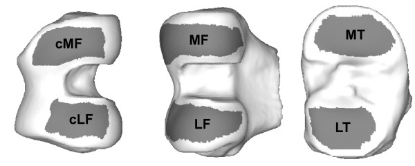
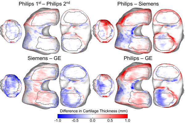
Twelve subjects with symptoms of knee OA and one or more risk factors had their symptomatic knee scanned on each of the three vendor's scanners located in three sites in the UK: Manchester (Philips), York (GE), and Liverpool (Siemens). The NIH OAI study protocol was used for the Siemens scanner, and equivalent protocols were developed for the Philips and GE scanners with vendors' advice. Cartilage was segmented manually from sagittal 3D images. By using recently described techniques for Anatomically Corresponded Regional Analysis of Cartilage (ACRAC), a statistical model was used anatomically to align all the images and to produce detailed maps of mean differences in cartilage-thickness measures between scanners. Measures of cartilage mean thickness were computed in anatomically equivalent regions for each subject and scanner image.

RESULTS

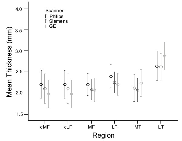
The ranges of mean cartilage-thickness measures for this cohort were similar for all regions and across all scanners. Philips intrascanner root-mean-square coefficients of variation were low in the range from 2.6% to 4.6%. No significant differences were found for thickness measures of the weight-bearing femorotibial regions from the Philips and Siemens images except for the central medial femur compartment (P = 0.04). Compared with the other two scanners, the GE scanner provided consistently lower mean thickness measures in the central femoral regions (mean difference, -0.16 mm) and higher measures in the tibial compartments (mean difference, +0.19 mm).

CONCLUSIONS

The OAI knee-imaging protocol, developed on the Siemens platform, can be applied to research and trials by using other vendors' 3T scanners giving comparable morphologic results. Accurate sequence optimization, differences in image postprocessing, and extremity coil type are critical factors for interscanner precision of quantitative analysis of cartilage morphology. It is still recommended that longitudinal observations on individuals should be performed on the same scanner and that assessment of intra- and interscanner precision errors is undertaken before commencement of the main study.

摘要

简介

磁共振成像(MRI)中的软骨厚度已被确定为膝关节骨关节炎(OA)研究中的一个潜在生物标志物。通过使用不同供应商的扫描仪在多个中心获取 MRI 数据,将有助于招募患者并缩短 OA 试验的持续时间。有几家供应商生产 3T 的 MRI 扫描仪,包括西门子、飞利浦医疗系统和通用电气医疗集团。本研究旨在调查来自三个不同供应商的扫描仪之间,软骨形态的定量 MRI 评估是否具有可比性。

方法

12 名有膝关节 OA 症状且有一个或多个危险因素的患者,在英国三个地点的三台不同供应商的 MRI 扫描仪上进行了膝关节扫描:曼彻斯特(飞利浦)、约克(GE)和利物浦(西门子)。西门子扫描仪使用 NIH OAI 研究方案,根据供应商的建议,为飞利浦和 GE 扫描仪制定了等效方案。通过使用最近描述的解剖对应区域软骨分析(ACRAC)技术,使用统计学模型对所有图像进行解剖对齐,并生成扫描仪间软骨厚度测量值差异的详细图谱。为每个患者和扫描仪图像计算了解剖学上等效区域的软骨平均厚度测量值。

结果

该队列的所有区域和所有扫描仪的平均软骨厚度测量值范围相似。飞利浦扫描仪的内部扫描均方根变异系数在 2.6%至 4.6%之间较低。除了中央内侧股骨间室(P=0.04),飞利浦和西门子图像的负重股骨胫骨区域的厚度测量值没有发现显著差异。与其他两台扫描仪相比,GE 扫描仪在中央股骨区域提供的平均厚度测量值始终较低(平均差异为-0.16 毫米),在胫骨区域的测量值较高(平均差异为+0.19 毫米)。

结论

基于西门子平台开发的 OAI 膝关节成像方案,可应用于使用其他供应商 3T 扫描仪进行的研究和试验,提供可比的形态学结果。准确的序列优化、图像后处理差异和肢体线圈类型是定量分析软骨形态的扫描仪间精度的关键因素。仍然建议对个体进行纵向观察时,应在同一台扫描仪上进行,并且在进行主要研究之前,应进行扫描仪内和扫描仪间精度误差的评估。

https://cdn.ncbi.nlm.nih.gov/pmc/blobs/5e46/2991039/88a6e26a4503/ar3174-3.jpg
https://cdn.ncbi.nlm.nih.gov/pmc/blobs/5e46/2991039/94cd6595976e/ar3174-1.jpg
https://cdn.ncbi.nlm.nih.gov/pmc/blobs/5e46/2991039/02cf7d6a5e41/ar3174-2.jpg
https://cdn.ncbi.nlm.nih.gov/pmc/blobs/5e46/2991039/88a6e26a4503/ar3174-3.jpg
https://cdn.ncbi.nlm.nih.gov/pmc/blobs/5e46/2991039/94cd6595976e/ar3174-1.jpg
https://cdn.ncbi.nlm.nih.gov/pmc/blobs/5e46/2991039/02cf7d6a5e41/ar3174-2.jpg
https://cdn.ncbi.nlm.nih.gov/pmc/blobs/5e46/2991039/88a6e26a4503/ar3174-3.jpg

相似文献

1
Comparison of 3T MR scanners in regional cartilage-thickness analysis in osteoarthritis: a cross-sectional multicenter, multivendor study.3T MR 扫描仪在骨关节炎区域性软骨厚度分析中的比较:一项横断面多中心、多厂商研究。
Arthritis Res Ther. 2010;12(5):R202. doi: 10.1186/ar3174. Epub 2010 Oct 28.
2
Magnetic resonance transverse relaxation time T2 of knee cartilage in osteoarthritis at 3-T: a cross-sectional multicentre, multivendor reproducibility study.3T 下骨关节炎膝关节软骨磁共振横向弛豫时间 T2:一项多中心、多厂家的横断面研究。
Skeletal Radiol. 2013 Apr;42(4):511-20. doi: 10.1007/s00256-012-1511-5. Epub 2012 Sep 28.
3
Precision of 3.0 Tesla quantitative magnetic resonance imaging of cartilage morphology in a multicentre clinical trial.多中心临床试验中3.0特斯拉定量磁共振成像对软骨形态的精准度
Ann Rheum Dis. 2008 Dec;67(12):1683-8. doi: 10.1136/ard.2007.076919. Epub 2008 Feb 18.
4
Can cartilage loss be detected in knee osteoarthritis (OA) patients with 3-6 months' observation using advanced image analysis of 3T MRI?在 3T MRI 高级图像分析的 3-6 个月观察下,能否检测到膝骨关节炎(OA)患者的软骨损失?
Osteoarthritis Cartilage. 2010 May;18(5):677-83. doi: 10.1016/j.joca.2010.02.010. Epub 2010 Feb 26.
5
Anatomically corresponded regional analysis of cartilage in asymptomatic and osteoarthritic knees by statistical shape modelling of the bone.基于骨的统计形状建模对无症状和骨关节炎膝关节软骨进行解剖对应区域分析。
IEEE Trans Med Imaging. 2010 Aug;29(8):1541-59. doi: 10.1109/TMI.2010.2047653. Epub 2010 Apr 8.
6
Automated techniques for visualization and mapping of articular cartilage in MR images of the osteoarthritic knee: a base technique for the assessment of microdamage and submicro damage.骨关节炎膝关节磁共振成像中关节软骨可视化与映射的自动化技术:一种评估微损伤和亚微损伤的基础技术。
IEEE Trans Nanobioscience. 2002 Mar;1(1):42-51. doi: 10.1109/tnb.2002.806916.
7
The Osteoarthritis Initiative (OAI) magnetic resonance imaging quality assurance update.《骨关节炎倡议(OAI)磁共振成像质量保证更新》。
Osteoarthritis Cartilage. 2013 Jan;21(1):110-6. doi: 10.1016/j.joca.2012.10.011. Epub 2012 Oct 23.
8
Magnetic resonance image segmentation using semi-automated software for quantification of knee articular cartilage---initial evaluation of a technique for paired scans.使用半自动软件进行磁共振图像分割以定量膝关节软骨——配对扫描技术的初步评估
Skeletal Radiol. 2009 May;38(5):505-11. doi: 10.1007/s00256-009-0658-1. Epub 2009 Feb 28.
9
Relationship between trabecular bone structure and articular cartilage morphology and relaxation times in early OA of the knee joint using parallel MRI at 3 T.使用3T并行磁共振成像研究膝关节早期骨关节炎中小梁骨结构与关节软骨形态及弛豫时间的关系。
Osteoarthritis Cartilage. 2008 Oct;16(10):1150-9. doi: 10.1016/j.joca.2008.02.018. Epub 2008 Apr 2.
10
Region of interest analysis: by selecting regions with denuded areas can we detect greater amounts of change?感兴趣区域分析:通过选择有裸露区域的区域,我们能否检测到更多的变化?
Osteoarthritis Cartilage. 2010 Feb;18(2):175-83. doi: 10.1016/j.joca.2009.08.002. Epub 2009 Aug 29.

引用本文的文献

1
DADP: Dynamic abnormality detection and progression for longitudinal knee magnetic resonance images from the Osteoarthritis Initiative.DADP:骨关节炎倡议组织纵向膝关节磁共振图像的动态异常检测与进展分析
Med Image Anal. 2022 Apr;77:102343. doi: 10.1016/j.media.2021.102343. Epub 2022 Jan 1.
2
The International Workshop on Osteoarthritis Imaging Knee MRI Segmentation Challenge: A Multi-Institute Evaluation and Analysis Framework on a Standardized Dataset.骨关节炎影像学膝关节MRI分割挑战赛国际研讨会:基于标准化数据集的多机构评估与分析框架
Radiol Artif Intell. 2021 Feb 10;3(3):e200078. doi: 10.1148/ryai.2021200078. eCollection 2021 May.
3

本文引用的文献

1
Measurement and visualisation of focal cartilage thickness change by MRI in a study of knee osteoarthritis using a novel image analysis tool.使用一种新的图像分析工具,对膝关节骨关节炎进行 MRI 测量和可视化研究,以观察焦点软骨厚度的变化。
Br J Radiol. 2010 Nov;83(995):940-8. doi: 10.1259/bjr/68875123. Epub 2010 Mar 11.
2
Regional analysis of femorotibial cartilage loss in a subsample from the Osteoarthritis Initiative progression subcohort.骨关节炎倡议进展亚队列中一个子样本的股胫关节软骨损失区域分析。
Osteoarthritis Cartilage. 2009 Mar;17(3):291-7. doi: 10.1016/j.joca.2008.07.008. Epub 2008 Sep 11.
3
One year change of knee cartilage morphology in the first release of participants from the Osteoarthritis Initiative progression subcohort: association with sex, body mass index, symptoms and radiographic osteoarthritis status.
Use of acoustic emission to identify novel candidate biomarkers for knee osteoarthritis (OA).
利用声发射技术鉴定膝关节骨关节炎(OA)的新型候选生物标志物。
PLoS One. 2019 Oct 16;14(10):e0223711. doi: 10.1371/journal.pone.0223711. eCollection 2019.
4
Learning-Based Cost Functions for 3-D and 4-D Multi-Surface Multi-Object Segmentation of Knee MRI: Data From the Osteoarthritis Initiative.基于学习的膝关节 MRI 三维和四维多表面多目标分割成本函数:来自骨关节炎倡议的数据。
IEEE Trans Med Imaging. 2018 May;37(5):1103-1113. doi: 10.1109/TMI.2017.2781541.
5
Workflow assessing the effect of gait alterations on stresses in the medial tibial cartilage - combined musculoskeletal modelling and finite element analysis.评估步态改变对内侧胫骨软骨内应力影响的工作流程 - 结合肌肉骨骼建模和有限元分析。
Sci Rep. 2017 Dec 12;7(1):17396. doi: 10.1038/s41598-017-17228-x.
6
OARSI Clinical Trials Recommendations: Hip imaging in clinical trials in osteoarthritis.骨关节炎研究学会国际联盟(OARSI)临床试验建议:骨关节炎临床试验中的髋关节成像
Osteoarthritis Cartilage. 2015 May;23(5):716-31. doi: 10.1016/j.joca.2015.03.004.
7
Imaging of cartilage and bone: promises and pitfalls in clinical trials of osteoarthritis.软骨与骨的成像:骨关节炎临床试验中的前景与陷阱
Osteoarthritis Cartilage. 2014 Oct;22(10):1516-32. doi: 10.1016/j.joca.2014.06.023.
8
No damage of joint cartilage of the lower limbs in an ultra-endurance athlete--an MRI-study.超耐力运动员下肢关节软骨无损伤——一项 MRI 研究。
BMC Musculoskelet Disord. 2013 Dec 5;14:343. doi: 10.1186/1471-2474-14-343.
9
Impact of different coils on biochemical T2 and T2* relaxation time mapping of articular patella cartilage.不同线圈对髌股关节软骨生化 T2 和 T2*弛豫时间成像的影响。
Skeletal Radiol. 2013 Nov;42(11):1565-72. doi: 10.1007/s00256-013-1699-z. Epub 2013 Aug 23.
10
Magnetic resonance transverse relaxation time T2 of knee cartilage in osteoarthritis at 3-T: a cross-sectional multicentre, multivendor reproducibility study.3T 下骨关节炎膝关节软骨磁共振横向弛豫时间 T2:一项多中心、多厂家的横断面研究。
Skeletal Radiol. 2013 Apr;42(4):511-20. doi: 10.1007/s00256-012-1511-5. Epub 2012 Sep 28.
骨关节炎倡议进展亚队列首批参与者膝关节软骨形态的一年变化:与性别、体重指数、症状及影像学骨关节炎状态的关联
Ann Rheum Dis. 2009 May;68(5):674-9. doi: 10.1136/ard.2008.089904. Epub 2008 Jun 2.
4
Precision of 3.0 Tesla quantitative magnetic resonance imaging of cartilage morphology in a multicentre clinical trial.多中心临床试验中3.0特斯拉定量磁共振成像对软骨形态的精准度
Ann Rheum Dis. 2008 Dec;67(12):1683-8. doi: 10.1136/ard.2007.076919. Epub 2008 Feb 18.
5
Proposal for a nomenclature for magnetic resonance imaging based measures of articular cartilage in osteoarthritis.骨关节炎中基于磁共振成像的关节软骨测量术语命名提案。
Osteoarthritis Cartilage. 2006 Oct;14(10):974-83. doi: 10.1016/j.joca.2006.03.005. Epub 2006 May 26.
6
Comparison of quantitative cartilage measurements acquired on two 3.0T MRI systems from different manufacturers.在两台不同制造商的3.0T磁共振成像(MRI)系统上获得的定量软骨测量结果的比较。
J Magn Reson Imaging. 2006 May;23(5):770-3. doi: 10.1002/jmri.20561.
7
Accuracy and precision of quantitative assessment of cartilage morphology by magnetic resonance imaging at 3.0T.3.0T磁共振成像对软骨形态进行定量评估的准确性和精确性
Arthritis Rheum. 2005 Oct;52(10):3132-6. doi: 10.1002/art.21348.
8
Whole-Organ Magnetic Resonance Imaging Score (WORMS) of the knee in osteoarthritis.骨关节炎膝关节全器官磁共振成像评分(WORMS)
Osteoarthritis Cartilage. 2004 Mar;12(3):177-90. doi: 10.1016/j.joca.2003.11.003.
9
Femoro-tibial cartilage metrics from coronal MR image data: Technique, test-retest reproducibility, and findings in osteoarthritis.来自冠状面磁共振成像数据的股骨-胫骨软骨测量指标:技术、重测信度及骨关节炎的研究结果
Magn Reson Med. 2003 Dec;50(6):1229-36. doi: 10.1002/mrm.10648.
10
Magnetic resonance imaging measurement of knee cartilage volume in a multicentre study.多中心研究中膝关节软骨体积的磁共振成像测量
Rheumatology (Oxford). 2004 Jan;43(1):19-21. doi: 10.1093/rheumatology/keg426. Epub 2003 Aug 15.