Laboratory of Epidemiology, Demography and Biometry, of the National Institutes on Aging (NIH), Bethesda, MD, USA.
Neurobiol Aging. 2012 Feb;33(2):424.e1-10. doi: 10.1016/j.neurobiolaging.2010.09.027. Epub 2010 Nov 13.
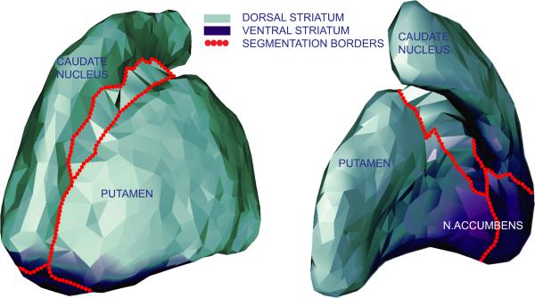
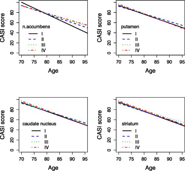
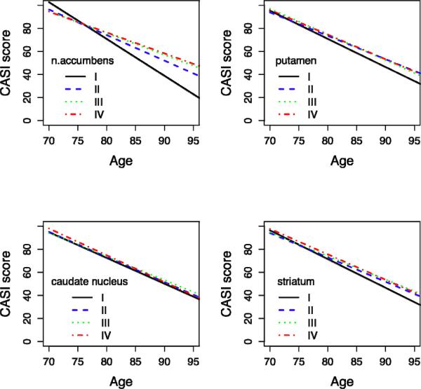
Striatal degeneration may contribute to cognitive impairment in older people. Here, we examine the relation of degeneration of the striatum and substructures to cognitive decline and dementia in subjects with a wide range of cognitive function. Data are from the prospective community-based Honolulu Asia Aging Study of Japanese American men born 1900-1919. Brain magnetic resonance imaging (MRI) (1.5 T) was acquired on a stratified subsample (n = 477) that included four groups defined by cognitive status relative to the scan date: subjects without dementia (n = 347), subjects identified as demented 2-3 years before brain scanning (n = 30), at the time of scanning (n = 58), and 3-5 years after scanning (n = 42). Volumes of the striatum, including the accumbens, putamen, and caudate nucleus were automatically estimated from T1 MR images. Global cognitive function was measured with the cognitive ability screening instrument (CASI), at four examinations spanning an 8-year interval. Trajectories of cognitive decline were estimated for each quartile of striatal volume using mixed models, controlling for demographic variables, measures of cerebro-vascular damage, global brain atrophy, and hippocampal volume. Diagnosis of dementia before, during, and after brain scanning was associated with smaller volumes of n. accumbens and putamen, but not with caudate nucleus volume. Subjects in the lowest quartile of n. accumbens volume, both in the total sample and in the subjects not diagnosed with dementia during the study, had a significantly (p < 0.0001) steeper decline in cognitive performance compared with those in the highest quartile. In conclusion, volumes of the n. accumbens and putamen are closely associated with the occurrence of dementia and n. accumbens volume predicts cognitive decline in older people. These associations were found independent of the magnitude of other pivotal markers of cognitive decline, i.e. cerebro-vascular damage and hippocampal volume. The present study suggests a role for the ventral striatum in the development of clinical dementia.
纹状体退化可能导致老年人认知障碍。在这里,我们研究了纹状体和亚结构的退化与认知功能广泛的受试者认知能力下降和痴呆之间的关系。数据来自前瞻性基于社区的日本裔美国男性 Honolulu Asia Aging 研究,这些男性出生于 1900-1919 年。对分层的子样本(n=477)进行了脑磁共振成像(MRI)(1.5T)采集,该子样本包括根据相对于扫描日期的认知状态定义的四个组:无痴呆症的受试者(n=347)、在扫描前 2-3 年被诊断为痴呆症的受试者(n=30)、在扫描时(n=58)和扫描后 3-5 年(n=42)的受试者。使用 T1 MR 图像自动估计纹状体(包括伏隔核)、壳核和尾状核的体积。使用认知能力筛查工具(CASI)在跨越 8 年的 4 次检查中测量整体认知功能。使用混合模型为每个纹状体体积四分位数估计认知下降轨迹,控制人口统计学变量、脑血管损伤、整体脑萎缩和海马体积的测量值。在扫描前、扫描期间和扫描后诊断为痴呆症与 n. 伏隔核和壳核的体积较小有关,但与尾状核体积无关。在总样本和在研究期间未被诊断为痴呆症的受试者中,n. 伏隔核体积最低四分位数的受试者认知表现下降明显更为陡峭(p<0.0001)。总之,n. 伏隔核和壳核的体积与痴呆症的发生密切相关,n. 伏隔核体积预测老年人的认知能力下降。这些关联是在排除其他认知能力下降的关键标志物(即脑血管损伤和海马体积)的大小后发现的。本研究表明腹侧纹状体在临床痴呆的发展中起作用。