Polgenix, Cleveland, Ohio, USA.
Nat Med. 2010 Dec;16(12):1444-9. doi: 10.1038/nm.2260. Epub 2010 Nov 14.
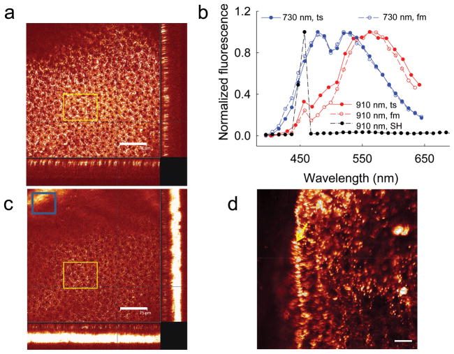
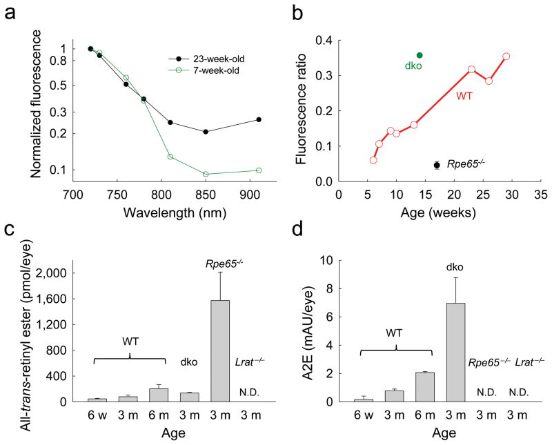
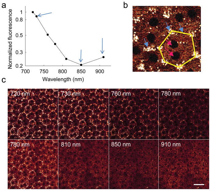
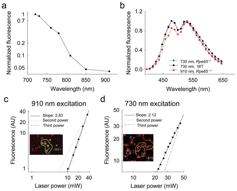
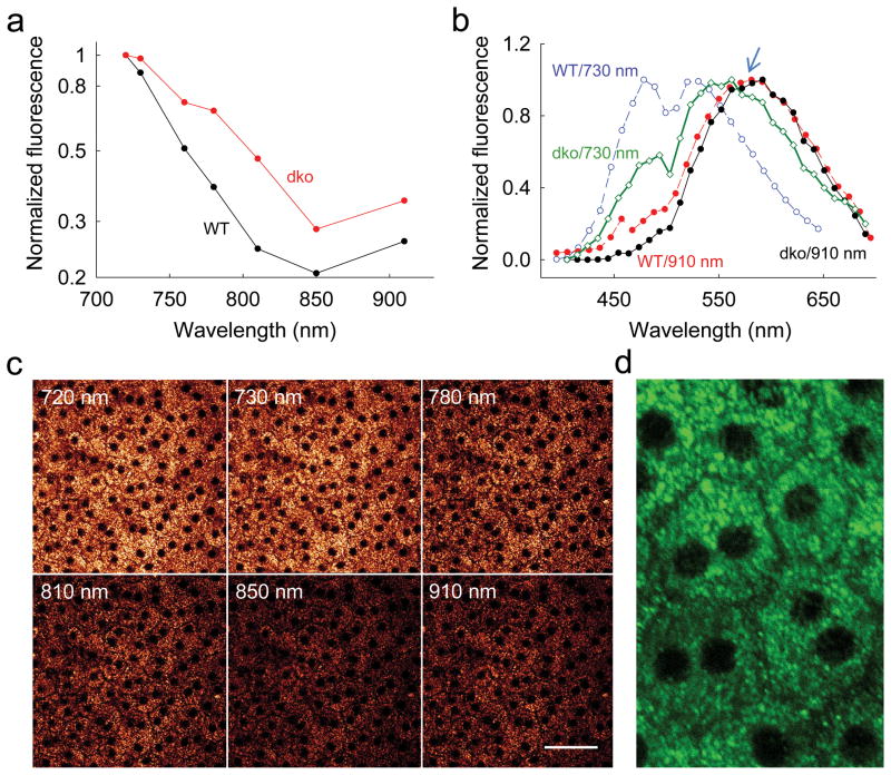
Multiphoton excitation fluorescence microscopy (MPM) can image certain molecular processes in vivo. In the eye, fluorescent retinyl esters in subcellular structures called retinosomes mediate regeneration of the visual chromophore, 11-cis-retinal, by the visual cycle. But harmful fluorescent condensation products of retinoids also occur in the retina. We report that in wild-type mice, excitation with a wavelength of ∼730 nm identified retinosomes in the retinal pigment epithelium, and excitation with a wavelength of ∼910 nm revealed at least one additional retinal fluorophore. The latter fluorescence was absent in eyes of genetically modified mice lacking a functional visual cycle, but accentuated in eyes of older wild-type mice and mice with defective clearance of all-trans-retinal, an intermediate in the visual cycle. MPM, a noninvasive imaging modality that facilitates concurrent monitoring of retinosomes along with potentially harmful products in aging eyes, has the potential to detect early molecular changes due to age-related macular degeneration and other defects in retinoid metabolism.
多光子激发荧光显微镜(MPM)可用于活体成像某些分子过程。在眼睛中,称为视脂体的亚细胞结构中的荧光视黄酯通过视觉循环介导视觉色素 11-顺式视黄醛的再生。但是,视黄醇的有害荧光缩合产物也会出现在视网膜中。我们报告说,在野生型小鼠中,用约 730nm 的波长激发可以识别视网膜色素上皮中的视脂体,而用约 910nm 的波长激发可以揭示至少一种额外的视网膜荧光团。在缺乏功能性视觉循环的基因修饰小鼠的眼睛中,后者的荧光缺失,但在年龄较大的野生型小鼠和全反式视黄醛清除有缺陷的小鼠的眼睛中,这种荧光增强,全反式视黄醛是视觉循环中的一种中间产物。MPM 是一种非侵入性的成像方式,可促进同时监测衰老眼中的视脂体以及潜在的有害产物,它有可能检测到与年龄相关的黄斑变性和其他视黄醇代谢缺陷相关的早期分子变化。