Chen Maxine M, Lee Chia-Yao, Leland Hyuma A, Silletti Steve
Moores Cancer Center, University of California, San Diego, La Jolla, CA 92093, USA.
Tumour Biol. 2011 Apr;32(2):347-57. doi: 10.1007/s13277-010-0127-4. Epub 2010 Nov 16.
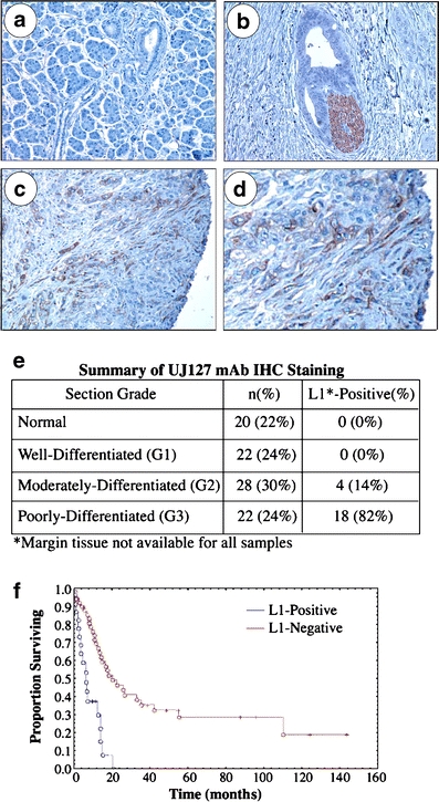
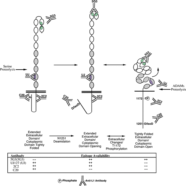
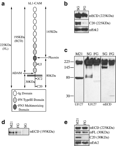
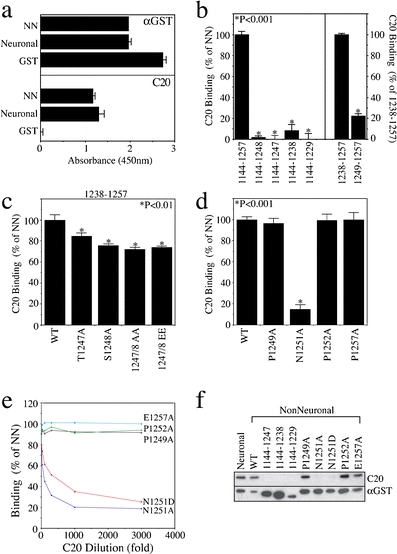
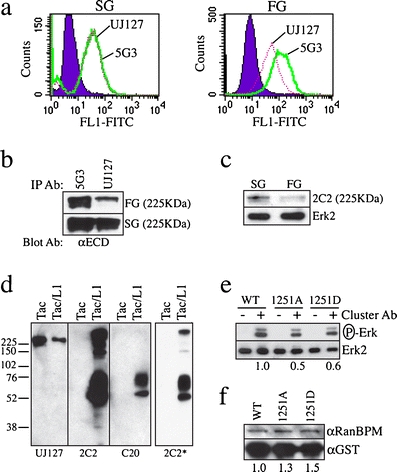
The neural cell adhesion molecule L1 has recently been shown to be expressed in pancreatic adenocarcinoma (PDAC) cells. In this report, we demonstrate that L1 is expressed by moderately- to poorly-differentiated PDAC cells in situ, and that L1 expression is a predictor of poor patient survival. In vitro, reduced reactivity of an anti-L1 carboxy-terminus-specific antibody was observed in the more poorly differentiated fast-growing (FG) variant of the COLO357 population, versus its well-differentiated slow-growing (SG) counterpart, even though they express equivalent total L1. The carboxy-terminus of L1 mediates binding to the MAP kinase-regulating protein RanBPM and mutation of T1247/S1248 within this region attenuates the expression of malignancy associated proteins and L1-induced tumorigenicity in mice. Therefore, we reasoned that the differential epitope exposure observed might be indicative of modifications responsible for regulating these events. However, epitope mapping demonstrated that the major determinant of binding was actually N1251; mutation of T1247 and S1248, alone or together, had little effect on C20 binding. Moreover, cluster assays using CD25 ectodomain/L1 cytoplasmic domain chimeras demonstrated the N1251-dependent, RanBPM-independent stimulation of erk phosphorylation in these cells. Reactivity of this antibody also reflects the differential exposure of extracellular epitopes in these COLO357 sublines, consistent with the previous demonstration of L1 ectodomain conformation modulation by intracellular modifications. These data further support a central role for L1 in PDAC, and define a specific role for carboxy-terminal residues including N1251 in the regulation of L1 activity in PDAC cells.
神经细胞黏附分子L1最近被证明在胰腺腺癌(PDAC)细胞中表达。在本报告中,我们证明L1在原位中、低分化的PDAC细胞中表达,并且L1表达是患者预后不良的一个预测指标。在体外,抗L1羧基末端特异性抗体在COLO357群体中分化较差、生长较快(FG)的变体中反应性降低,与其分化良好、生长较慢(SG)的对应变体相比,尽管它们表达的总L1相当。L1的羧基末端介导与丝裂原活化蛋白激酶调节蛋白RanBPM的结合,该区域内T1247/S1248的突变会减弱恶性相关蛋白的表达以及L1诱导的小鼠致瘤性。因此,我们推断观察到的差异表位暴露可能指示负责调节这些事件的修饰。然而,表位作图表明结合的主要决定因素实际上是N1251;T1247和S1248单独或共同突变对C20结合几乎没有影响。此外,使用CD25胞外域/L1胞质域嵌合体的聚类分析表明,在这些细胞中存在N1251依赖性、RanBPM非依赖性的erk磷酸化刺激。该抗体的反应性还反映了这些COLO357亚系中细胞外表位的差异暴露,这与先前关于细胞内修饰对L1胞外域构象调节的证明一致。这些数据进一步支持了L1在PDAC中的核心作用,并确定了包括N1251在内的羧基末端残基在调节PDAC细胞中L1活性方面的特定作用。