Medizinische Klinik III, Universitätsklinikum Carl Gustav Carus Dresden, Fetscherstraße 74, 01307 Dresden, Germany.
Klinik und Poliklinik für Viszeral-, Thorax- und Gefäßchirurgie, Universitätsklinikum Carl Gustav Carus Dresden, Fetscherstraße 74, 01307 Dresden, Germany.
Int J Mol Sci. 2019 Jul 5;20(13):3305. doi: 10.3390/ijms20133305.
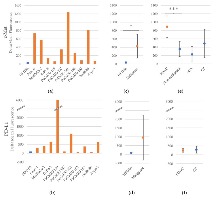
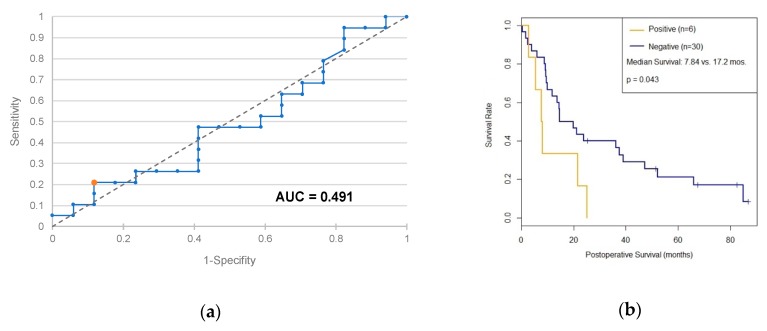
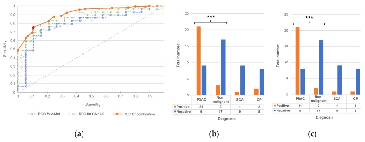
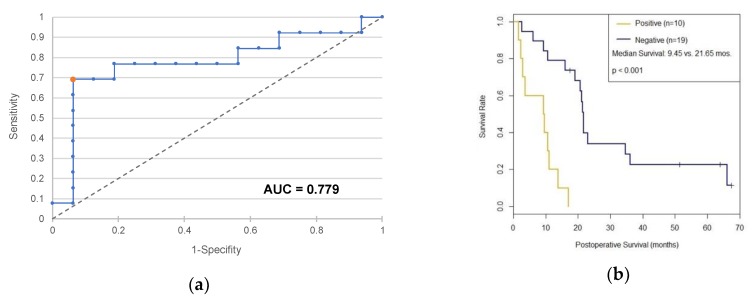
Exosomes are membrane vesicles which offer potential as blood derived biomarkers for malign tumors in clinical practice. Pancreatic cancer is counted among cancer diseases with the highest mortality. The present work seeks to assess whether pancreatic carcinomas release exosomes which express c-Met (proto-oncogene mesenchymal-epithelial transition factor) and PD-L1 (programmed cell death 1 ligand 1), and whether the detection of such expression in serum has diagnostic or prognostic meaning for the affected patients. Exosome isolation was performed on culture media of one benign pancreatic cell line and ten pancreatic carcinoma cell lines as well as on serum samples from 55 patients with pancreatic ductal adenocarcinoma (PDAC), 26 patients with chronic pancreatitis and 10 patients with benign serous cyst adenoma of the pancreas. Exosomes were bound to latex beads and stained with antibodies against c-Met or PD-L1. Analysis of fluorescence intensity was performed by flow cytometry. In terms of c-Met, the mean fluorescence intensity of PDAC-patients was significantly higher than the fluorescence intensity of the comparative patients with benign disease ( < 0.001). A diagnostic test based on c-Met resulted in a sensitivity of 70%, a specificity of 85% and a diagnostic odds ratio of 13:2. The specificity of the test can be further improved by combining it with the established tumor marker carbohydrate antigen 19-9 (CA 19-9). In addition, c-Met-positive patients showed a significantly shorter postoperative survival time (9.5 vs. 21.7 months, < 0.001). In terms of PD-L1, no significant difference between fluorescence intensity of PDAC-patients and comparative patients was detectable. However, PD-L1-positive PDAC-patients also showed a significantly shorter postoperative survival time (7.8 vs. 17.2 months, = 0.043). Thus, both markers can be considered as negative prognostic factors.
外泌体是一种膜囊泡,具有作为临床恶性肿瘤血液来源生物标志物的潜力。胰腺癌是癌症死亡率最高的疾病之一。本研究旨在评估胰腺癌细胞是否释放表达 c-Met(原癌基因间质上皮转化因子)和 PD-L1(程序性细胞死亡 1 配体 1)的外泌体,以及在血清中检测到这种表达对受影响患者是否具有诊断或预后意义。在外泌体分离过程中,使用了一种良性胰腺细胞系和十种胰腺癌细胞系的培养基以及 55 例胰腺导管腺癌(PDAC)患者、26 例慢性胰腺炎患者和 10 例良性浆液性囊腺瘤患者的血清样本。外泌体与乳胶珠结合,并使用针对 c-Met 或 PD-L1 的抗体进行染色。通过流式细胞术分析荧光强度。在 c-Met 方面,PDAC 患者的平均荧光强度明显高于良性疾病患者的荧光强度(<0.001)。基于 c-Met 的诊断测试的灵敏度为 70%,特异性为 85%,诊断优势比为 13:2。通过将该测试与已建立的肿瘤标志物碳水化合物抗原 19-9(CA 19-9)结合,可以进一步提高该测试的特异性。此外,c-Met 阳性患者的术后生存时间明显缩短(9.5 个月与 21.7 个月,<0.001)。在 PD-L1 方面,PDAC 患者和对照组患者的荧光强度之间没有检测到显著差异。然而,PD-L1 阳性 PDAC 患者的术后生存时间也明显缩短(7.8 个月与 17.2 个月,=0.043)。因此,这两个标志物都可以被认为是负预后因素。