Lin William, Kang Un Jung
Department of Neurology, University of Chicago Medical Center, Chicago, Illinois 60637, USA.
BMC Cell Biol. 2010 Nov 22;11:90. doi: 10.1186/1471-2121-11-90.
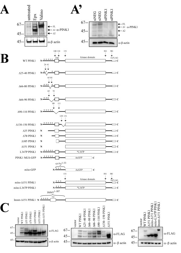
PINK1 is a mitochondria-targeted kinase that constitutively localizes to both the mitochondria and the cytosol. The mechanism of how PINK1 achieves cytosolic localization following mitochondrial processing remains unknown. Understanding PINK1 subcellular localization will give us insights into PINK1 functions and how mutations in PINK1 lead to Parkinson's disease. We asked how the mitochondrial localization signal, the transmembrane domain, and the kinase domain participate in PINK1 localization.
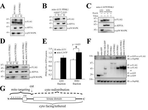
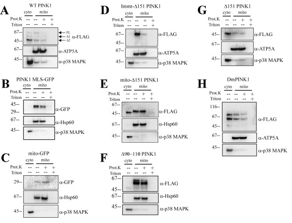
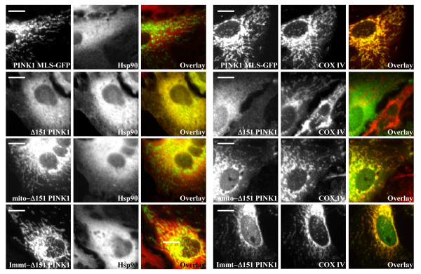
We confirmed that PINK1 mitochondrial targeting signal is responsible for mitochondrial localization. Once inside the mitochondria, we found that both PINK1 transmembrane and kinase domain are important for membrane tethering and cytosolic-facing topology. We also showed that PINK1 dual subcellular distribution requires both Hsp90 interaction with the kinase domain and the proteolysis at a cleavage site downstream of the transmembrane domain because removal of this cleavage site completely abolished cytosolic PINK1. In addition, the disruption of the Hsp90-PINK1 interaction increased mitochondrial PINK1 level.
Together, we believe that once PINK1 enters the mitochondria, PINK1 adopts a tethered topology because the transmembrane domain and the kinase domain prevent PINK1 forward movement into the mitochondria. Subsequent proteolysis downstream of the transmembrane domain then releases PINK1 for retrograde movement while PINK1 kinase domain interacts with Hsp90 chaperone. The significance of this dual localization could mean that PINK1 has compartmental-specific functions.
PINK1是一种定位于线粒体的激酶,其组成性地定位于线粒体和细胞质中。PINK1在线粒体加工后如何实现细胞质定位的机制尚不清楚。了解PINK1的亚细胞定位将有助于我们深入了解PINK1的功能以及PINK1突变如何导致帕金森病。我们研究了线粒体定位信号、跨膜结构域和激酶结构域如何参与PINK1的定位。
我们证实PINK1的线粒体靶向信号负责其在线粒体中的定位。一旦进入线粒体,我们发现PINK1的跨膜结构域和激酶结构域对于膜锚定和面向细胞质的拓扑结构都很重要。我们还表明,PINK1的双亚细胞分布需要Hsp90与激酶结构域的相互作用以及跨膜结构域下游切割位点的蛋白水解,因为去除该切割位点会完全消除细胞质中的PINK1。此外,Hsp90-PINK1相互作用的破坏会增加线粒体中PINK1的水平。
我们认为,一旦PINK1进入线粒体,它就会采用一种锚定的拓扑结构,因为跨膜结构域和激酶结构域会阻止PINK1向前移动到线粒体中。随后跨膜结构域下游的蛋白水解会释放PINK1以便逆行移动,同时PINK1激酶结构域与Hsp90伴侣相互作用。这种双定位的意义可能意味着PINK1具有特定区域的功能。