Department of Neurology, University of Texas Medical School at Houston, Houston, Texas 77030, USA.
BMC Neurosci. 2010 Nov 29;11:151. doi: 10.1186/1471-2202-11-151.
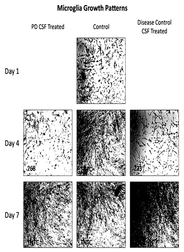
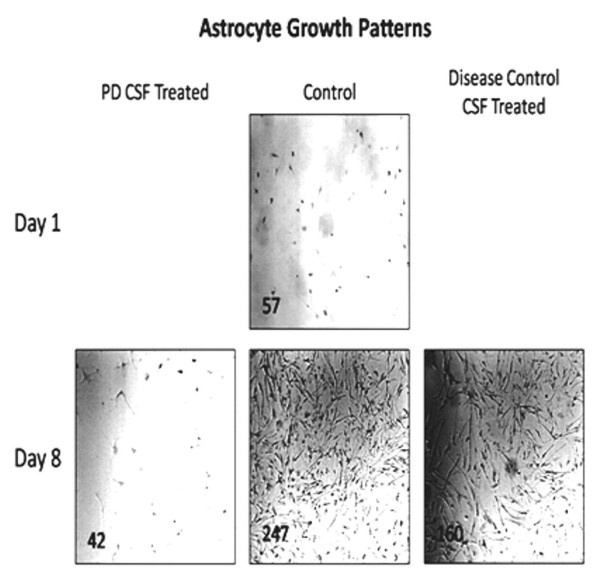
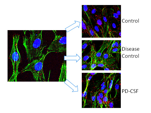
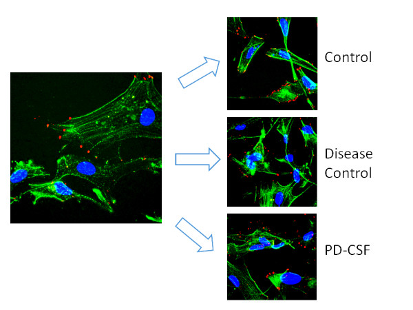
Excessive and abnormal accumulation of alpha-synuclein (α-synuclein) is a factor contributing to pathogenic cell death in Parkinson's disease. The purpose of this study, based on earlier observations of Parkinson's disease cerebrospinal fluid (PD-CSF) initiated cell death, was to determine the effects of CSF from PD patients on the functionally different microglia and astrocyte glial cell lines. Microglia cells from human glioblastoma and astrocytes from fetal brain tissue were cultured, grown to confluence, treated with fixed concentrations of PD-CSF, non-PD disease control CSF, or control no-CSF medium, then photographed and fluorescently probed for α-synuclein content by deconvolution fluorescence microscopy. Outcome measures included manually counted cell growth patterns from day 1-8; α-synuclein density and distribution by antibody tagged 3D model stacked deconvoluted fluorescent imaging.
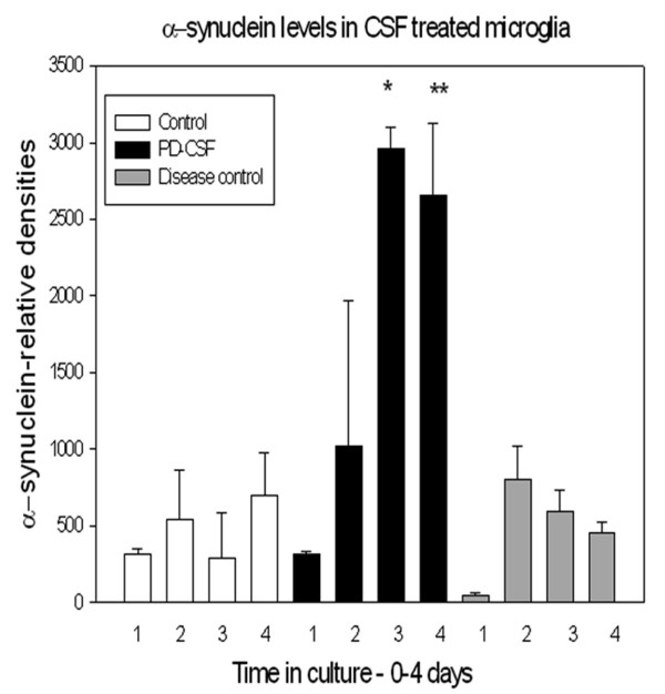
After PD-CSF treatment, microglia growth was reduced extensively, and a non-confluent pattern with morphological changes developed, that was not evident in disease control CSF and no-CSF treated cultures. Astrocyte growth rates were similarly reduced by exposure to PD-CSF, but morphological changes were not consistently noted. PD-CSF treated microglia showed a significant increase in α-synuclein content by day 4 compared to other treatments (p ≤ 0.02). In microglia only, α-synuclein aggregated and redistributed to peri-nuclear locations.
Cultured microglia and astrocytes are differentially affected by PD-CSF exposure compared to non-PD-CSF controls. PD-CSF dramatically impacts microglia cell growth, morphology, and α-synuclein deposition compared to astrocytes, supporting the hypothesis of cell specific susceptibility to PD-CSF toxicity.
α-突触核蛋白(α-synuclein)的过度和异常积累是导致帕金森病致病细胞死亡的一个因素。本研究基于帕金森病脑脊液(PD-CSF)引发细胞死亡的早期观察结果,旨在确定 PD 患者 CSF 对功能不同的小胶质细胞和星形胶质细胞系的影响。从小胶质细胞瘤中培养人类胶质母细胞瘤细胞和从胎脑组织中培养星形胶质细胞,使其达到汇合状态,用固定浓度的 PD-CSF、非 PD 疾病对照 CSF 或对照无 CSF 培养基处理,然后通过共焦荧光显微镜对 α-突触核蛋白含量进行荧光探测和反卷积。结果测量包括从第 1 天到第 8 天手动计数细胞生长模式;通过抗体标记的 3D 模型堆叠反卷积荧光成像测量 α-突触核蛋白的密度和分布。
PD-CSF 处理后,小胶质细胞的生长受到广泛抑制,形成非融合模式,并伴有形态变化,而在疾病对照 CSF 和无 CSF 处理的培养物中则不明显。暴露于 PD-CSF 也会导致星形胶质细胞生长速度降低,但形态变化并不明显。与其他处理相比,PD-CSF 处理的小胶质细胞在第 4 天表现出明显增加的 α-突触核蛋白含量(p≤0.02)。只有小胶质细胞中,α-突触核蛋白聚集并重新分布到核周位置。
与非 PD-CSF 对照相比,培养的小胶质细胞和星形胶质细胞对 PD-CSF 暴露的反应不同。与星形胶质细胞相比,PD-CSF 对小胶质细胞的生长、形态和 α-突触核蛋白沉积有显著影响,支持细胞对 PD-CSF 毒性的特定易感性假说。