Cardiovascular Division, Department of Medicine, University of Pennsylvania School of Medicine, Philadelphia, PA, USA.
Circ Arrhythm Electrophysiol. 2011 Feb;4(1):49-55. doi: 10.1161/CIRCEP.110.959957. Epub 2010 Dec 3.
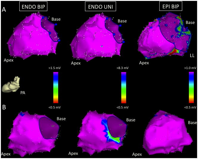
Patients with nonischemic left ventricular cardiomyopathy (LVCM) and ventricular tachycardia (Vt) have complex 3-dimensional substrate with variable involvement of the endocardium (ENDO) and epicardium (EPI). The purpose of this study was to determine whether ENDO unipolar (UNI) mapping with a larger electric field of view could identify EPI low bipolar (BIP) voltage regions in patients with LVCM undergoing Vt ablation.
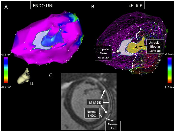
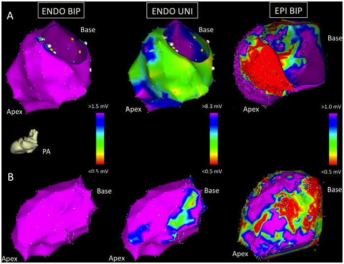
The reference value for normal ENDO unipolar voltage was determined from 6 patients without structural heart disease. Consecutive patients undergoing Vt ablation over an 8-year period with detailed (>100 points) LV ENDO and EPI mapping and normal LV ENDO BIP voltage were identified. From this cohort, we compared patients with structurally normal hearts and normal EPI BIP voltage (EPI-, group 1) with patients with LVCM and low LV EPI BIP voltage regions present (EPI+, group 2). Confluent regions of ENDO UNI and EPI BIP low voltage (>2 cm(2)) were measured. The normal signal amplitude was >8.27 mV for LV ENDO UNI electrograms. Detailed LV ENDO-EPI maps in 5 EPI- patients were compared with 11 EPI+ patients. Confluent ENDO UNI low-voltage regions were seen in 9 of 11 (82%) of the EPI+ (group 2) patients compared with none of 5 EPI- (group 1) patients (P<0.001). In all 9 patients with ENDO UNI low voltage, the ENDO UNI low-voltage regions were directly opposite to an area of EPI BIP low voltage (61% ENDO UNI-EPI BIP low-voltage area overlap).
EPI arrhythmia substrate can be reliably identified in most patients with LVCM using ENDO UNI voltage mapping in the absence of ENDO BIP abnormalities.
非缺血性左心室心肌病(LVCM)和室性心动过速(Vt)患者的 3 维基质复杂,心内膜(ENDO)和心外膜(EPI)的受累程度不同。本研究的目的是确定 LVCM 患者行 Vt 消融时,使用更大的电视野进行 ENDO 单极(UNI)标测是否可以识别 EPI 低双极(BIP)电压区。
从 6 例无结构性心脏病患者中确定了正常 ENDO 单极电压的参考值。连续 8 年期间,对接受 Vt 消融且具有详细(>100 个点)LV ENDO 和 EPI 标测且 LV ENDO BIP 电压正常的患者进行了识别。从该队列中,我们比较了结构正常且 EPI BIP 电压正常(EPI-,第 1 组)的患者与 LVCM 患者和存在低 LV EPI BIP 电压区的患者(EPI+,第 2 组)。测量了 ENDO UNI 和 EPI BIP 低电压(>2cm2)的融合区。LV ENDO UNI 电图的正常信号幅度>8.27mV。将 5 例 EPI-患者的详细 LV ENDO-EPI 图谱与 11 例 EPI+患者进行了比较。在 11 例 EPI+(第 2 组)患者中,有 9 例(82%)看到了融合的 ENDO UNI 低电压区,而在 5 例 EPI-(第 1 组)患者中无 1 例(P<0.001)。在所有 9 例 ENDO UNI 低电压的患者中,ENDO UNI 低电压区与 EPI BIP 低电压区直接相对(61%ENDO UNI-EPI BIP 低电压区重叠)。
在 LVCM 患者中,即使没有 ENDO BIP 异常,也可以使用 ENDO UNI 电压标测可靠地识别 EPI 心律失常基质。