• 文献检索
  • 文档翻译
  • 深度研究
  • 学术资讯
  • Suppr Zotero 插件Zotero 插件
  • 邀请有礼
  • 套餐&价格
  • 历史记录
应用&插件
Suppr Zotero 插件Zotero 插件浏览器插件Mac 客户端Windows 客户端微信小程序
定价
高级版会员购买积分包购买API积分包
服务
文献检索文档翻译深度研究API 文档MCP 服务
关于我们
关于 Suppr公司介绍联系我们用户协议隐私条款
关注我们

Suppr 超能文献

核心技术专利:CN118964589B侵权必究
粤ICP备2023148730 号-1Suppr @ 2026

文献检索

告别复杂PubMed语法,用中文像聊天一样搜索,搜遍4000万医学文献。AI智能推荐,让科研检索更轻松。

立即免费搜索

文件翻译

保留排版,准确专业,支持PDF/Word/PPT等文件格式,支持 12+语言互译。

免费翻译文档

深度研究

AI帮你快速写综述,25分钟生成高质量综述,智能提取关键信息,辅助科研写作。

立即免费体验

通过非侵入性光学分析诊断外阴病变:一项试点研究。

Diagnosis of vulvar lesions by non-invasive optical analysis: a pilot study.

作者信息

Vlastos Anne-Therese, Charvet Igor, Dellacasa Ilaria, Capanna Federica, Pelte Marie-Françoise, Thueler Philippe, Saint-Ghislain Michel, Depeursinge Christian, Meda Paolo

机构信息

Department of Gynecology and Obstetrics.

出版信息

Rare Tumors. 2009 Jul 22;1(1):e8. doi: 10.4081/rt.2009.e8.

DOI:10.4081/rt.2009.e8
PMID:21139902
原文链接:https://pmc.ncbi.nlm.nih.gov/articles/PMC2994441/
Abstract

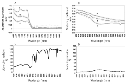
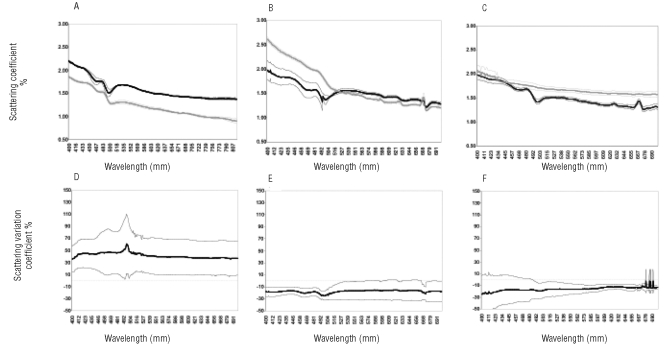
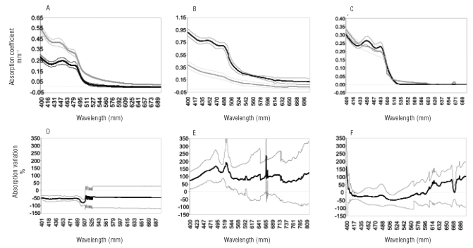
A procedure that could allow an early in vivo and non-invasive detection of vulvar lesions would be extremely useful. We tested an innovative optical method (Optiprobe), which uses a harmless, visible light source for the in vivo, on-line detection of minimal alterations in the structure of vulvar epithelium. A group of 3 female volunteers without gynecological symptoms were first screened to evaluate optical properties of normal vulvar tissue. Next, a group of 16 patients undergoing gynecological examination for vulvar lesions was evaluated by the Optiprobe at suspected sites before these sites were biopsied for histological analysis. Adjacent, non-involved sites were also measured to provide internal controls. Histological analysis of the biopsies identified one case that did not show obvious alterations, 4 cases of high-grade vulvar intraepithelial neoplasia (VIN), 5 cases of vulvitis, and 6 cases of lichen sclerosis (LS).The optical properties of the VIN cases were significantly different from those of controls, due to a decrease in the absorption spectra and an increase in the scattering spectra. In contrast, a significant increase in the absorption spectra and a decrease in the scattering spectra were observed in the cases of vulvitis. In the LS cases, the absorption spectra were as in controls, whereas the scattering spectra were significantly decreased. We conclude that the Optiprobe provides a useful tool for a rapid and non-invasive detection of vulvar alterations. The method should contribute to reduce the number of biopsies and to facilitate the long-term follow-up of vulvar lesions.

摘要

一种能够实现早期体内无创检测外阴病变的方法将非常有用。我们测试了一种创新的光学方法(Optiprobe),该方法使用无害的可见光源对体外的外阴上皮结构微小变化进行在线检测。首先对一组3名无妇科症状的女性志愿者进行筛查,以评估正常外阴组织的光学特性。接下来,对一组16名因外阴病变接受妇科检查的患者在疑似部位进行活检以进行组织学分析之前,先用Optiprobe进行评估。还对相邻的未受累部位进行测量以提供内部对照。活检的组织学分析发现1例未显示明显改变,4例高级别外阴上皮内瘤变(VIN),5例外阴炎和6例扁平苔藓(LS)。由于吸收光谱降低和散射光谱增加,VIN病例的光学特性与对照有显著差异。相比之下,外阴炎病例中观察到吸收光谱显著增加和散射光谱降低。在LS病例中,吸收光谱与对照相同,而散射光谱显著降低。我们得出结论,Optiprobe为快速无创检测外阴改变提供了一种有用的工具。该方法应有助于减少活检数量并便于对外阴病变进行长期随访。

https://cdn.ncbi.nlm.nih.gov/pmc/blobs/848a/2994441/39c5b36a21b8/rt-2009-1-e8-g003.jpg
https://cdn.ncbi.nlm.nih.gov/pmc/blobs/848a/2994441/8a7f1e4540c7/rt-2009-1-e8-g001.jpg
https://cdn.ncbi.nlm.nih.gov/pmc/blobs/848a/2994441/c3c1ee47b837/rt-2009-1-e8-g002.jpg
https://cdn.ncbi.nlm.nih.gov/pmc/blobs/848a/2994441/39c5b36a21b8/rt-2009-1-e8-g003.jpg
https://cdn.ncbi.nlm.nih.gov/pmc/blobs/848a/2994441/8a7f1e4540c7/rt-2009-1-e8-g001.jpg
https://cdn.ncbi.nlm.nih.gov/pmc/blobs/848a/2994441/c3c1ee47b837/rt-2009-1-e8-g002.jpg
https://cdn.ncbi.nlm.nih.gov/pmc/blobs/848a/2994441/39c5b36a21b8/rt-2009-1-e8-g003.jpg

相似文献

1
Diagnosis of vulvar lesions by non-invasive optical analysis: a pilot study.通过非侵入性光学分析诊断外阴病变:一项试点研究。
Rare Tumors. 2009 Jul 22;1(1):e8. doi: 10.4081/rt.2009.e8.
2
Histopathologic study of thin vulvar squamous cell carcinomas and associated cutaneous lesions: a correlative study of 48 tumors in 44 patients with analysis of adjacent vulvar intraepithelial neoplasia types and lichen sclerosus.薄型外阴鳞状细胞癌及相关皮肤病变的组织病理学研究:44例患者48个肿瘤的相关性研究,分析相邻外阴上皮内瘤变类型和硬化性苔藓。
Am J Surg Pathol. 2006 Mar;30(3):310-8. doi: 10.1097/01.pas.0000180444.71775.1a.
3
In the absence of (early) invasive carcinoma, vulvar intraepithelial neoplasia associated with lichen sclerosus is mainly of undifferentiated type: new insights in histology and aetiology.在无(早期)浸润性癌的情况下,与硬化性苔藓相关的外阴上皮内瘤变主要为未分化型:组织学和病因学的新见解。
J Clin Pathol. 2007 May;60(5):504-9. doi: 10.1136/jcp.2005.031989. Epub 2006 May 19.
4
Vulvar lichen sclerosus and squamous cell carcinoma: a cohort, case control, and investigational study with historical perspective; implications for chronic inflammation and sclerosis in the development of neoplasia.外阴硬化性苔藓与鳞状细胞癌:一项具有历史视角的队列研究、病例对照研究及调查研究;对肿瘤形成过程中慢性炎症和硬化的启示。
Hum Pathol. 1998 Sep;29(9):932-48. doi: 10.1016/s0046-8177(98)90198-8.
5
Vulvar intraepithelial neoplasia of the simplex (differentiated) type: a clinicopathologic study including analysis of HPV and p53 expression.单纯(分化型)外阴上皮内瘤变:一项包括人乳头瘤病毒(HPV)分析和p53表达的临床病理研究
Am J Surg Pathol. 2000 Mar;24(3):429-41. doi: 10.1097/00000478-200003000-00013.
6
Differentiated Vulvar Intraepithelial Neoplasia-like and Lichen Sclerosus-like Lesions in HPV-associated Squamous Cell Carcinomas of the Vulva.外阴 HPV 相关鳞状细胞癌中分化的外阴上皮内瘤样病变样和硬化性苔藓样病变。
Am J Surg Pathol. 2018 Jun;42(6):828-835. doi: 10.1097/PAS.0000000000001047.
7
Vulvar Lichen Sclerosus and Neoplastic Transformation: A Retrospective Study of 976 Cases.外阴硬化性苔藓与肿瘤转化:976例回顾性研究
J Low Genit Tract Dis. 2016 Apr;20(2):180-3. doi: 10.1097/LGT.0000000000000186.
8
[Review of precancerous vulvar lesions].[外阴癌前病变综述]
Cesk Patol. 2012 Jan;48(1):15-21.
9
Immunohistochemical expression of SOX2 in vulvar intraepithelial neoplasia and squamous cell carcinoma.SOX2 在外阴上皮内瘤变和鳞状细胞癌中的免疫组织化学表达。
Int J Gynecol Pathol. 2013 May;32(3):323-8. doi: 10.1097/PGP.0b013e31825d820e.
10
Lectins in the vulva. I. Normal vulvar epithelium and epithelium adjacent to vulvar intraepithelial neoplasia and squamous cell carcinoma.外阴中的凝集素。I. 正常外阴上皮以及与外阴上皮内瘤变和鳞状细胞癌相邻的上皮。
Int J Gynecol Pathol. 1998 Apr;17(2):154-61. doi: 10.1097/00004347-199804000-00010.

本文引用的文献

1
Elastic scattering spectroscopy as a diagnostic tool for differentiating pathologies in the gastrointestinal tract: preliminary testing.弹性散射光谱作为鉴别胃肠道病变的诊断工具:初步测试
J Biomed Opt. 1996 Apr;1(2):192-9. doi: 10.1117/12.231372.
2
Spatially resolved absolute diffuse reflectance measurements for noninvasive determination of the optical scattering and absorption coefficients of biological tissue.用于无创测定生物组织光学散射和吸收系数的空间分辨绝对漫反射测量。
Appl Opt. 1996 May 1;35(13):2304-14. doi: 10.1364/AO.35.002304.
3
Clinical determination of tissue optical properties by endoscopic spatially resolved reflectometry.
通过内镜空间分辨反射测量法进行组织光学特性的临床测定。
Appl Opt. 1996 Apr 1;35(10):1756-66. doi: 10.1364/AO.35.001756.
4
Diffuse reflectance spectroscopy of human adenomatous colon polyps in vivo.人腺瘤性结肠息肉的体内漫反射光谱分析
Appl Opt. 1999 Nov 1;38(31):6628-37. doi: 10.1364/ao.38.006628.
5
In vivo local determination of tissue optical properties: applications to human brain.组织光学特性的体内局部测定:在人脑中的应用
Appl Opt. 1999 Aug 1;38(22):4939-50. doi: 10.1364/ao.38.004939.
6
Diagnosis and grading of gastritis by non-invasive optical analysis.通过非侵入性光学分析诊断胃炎及对其进行分级
Eur J Gastroenterol Hepatol. 2004 Nov;16(11):1189-98. doi: 10.1097/00042737-200411000-00018.
7
In vivo endoscopic tissue diagnostics based on spectroscopic absorption, scattering, and phase function properties.
J Biomed Opt. 2003 Jul;8(3):495-503. doi: 10.1117/1.1578494.
8
A new optical method for the non-invasive detection of minimal tissue alterations.一种用于无创检测微小组织变化的新型光学方法。
Phys Med Biol. 2002 Jun 21;47(12):2095-108. doi: 10.1088/0031-9155/47/12/307.
9
Determination of optical parameters of human breast tissue from spatially resolved fluorescence: a diffusion theory model.
Appl Opt. 2002 Jul 1;41(19):4024-35. doi: 10.1364/ao.41.004024.
10
[Guidelines for diagnosis and therapy of intraepithelial neoplasia and early invasive carcinoma of the female lower genital system (cervix uteri, vagina, vulva) established by the AGK (Colposcopy Work Group in the OGGG [Austrian Society of Gynecology and Obstetrics])].[AGK(奥地利妇产科协会阴道镜工作组)制定的女性下生殖道系统(子宫颈、阴道、外阴)上皮内瘤变和早期浸润癌的诊断与治疗指南]
Gynakol Geburtshilfliche Rundsch. 2001;41(3):197-200. doi: 10.1159/000049475.