Institute of Clinical Biochemistry and Pathobiochemistry, German Diabetes Centre, Düsseldorf, Germany.
J Cell Mol Med. 2011 Nov;15(11):2399-410. doi: 10.1111/j.1582-4934.2010.01232.x.
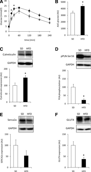
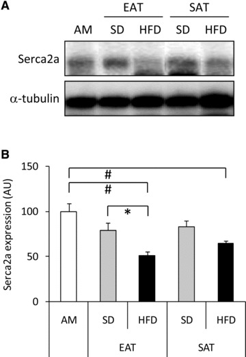
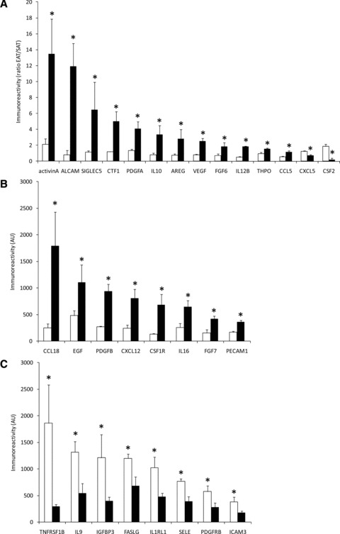
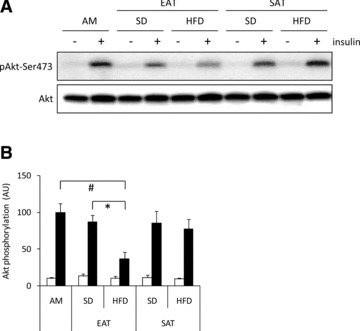
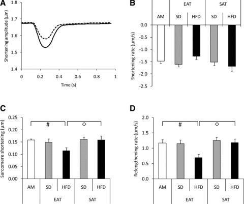
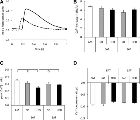
Epicardial adipose tissue (EAT) has been implicated in the development of heart disease. Nonetheless, the crosstalk between factors secreted from EAT and cardiomyocytes has not been studied. Here, we examined the effect of factors secreted from EAT on contractile function and insulin signalling in primary rat cardiomocytes. EAT and subcutaneous adipose tissue (SAT) were isolated from guinea pigs fed a high-fat (HFD) or standard diet. HFD feeding for 6 months induced glucose intolerance, and decreased fractional shortening and ejection fraction (all P < 0.05). Conditioned media (CM) generated from EAT and SAT explants were subjected to cytokine profiling using antibody arrays, or incubated with cardiomyocytes to assess the effects on insulin action and contractile function. Eleven factors were differentially secreted by EAT when compared to SAT. Furthermore, secretion of 30 factors by EAT was affected by HFD feeding. Most prominently, activin A-immunoreactivity was 6.4-fold higher in CM from HFD versus standard diet-fed animals and, 2-fold higher in EAT versus SAT. In cardiomyocytes, CM from EAT of HFD-fed animals increased SMAD2-phosphorylation, a marker for activin A-signalling, decreased sarcoplasmic-endoplasmic reticulum calcium ATPase 2a expression, and reduced insulin-mediated phosphorylation of Akt-Ser473 versus CM from SAT and standard diet-fed animals. Finally, CM from EAT of HFD-fed animals as compared to CM from the other groups markedly reduced sarcomere shortening and cytosolic Ca(2+) fluxes in cardiomyocytes. These data provide evidence for an interaction between factors secreted from EAT and cardiomyocyte function.
心外膜脂肪组织 (EAT) 与心脏病的发生发展有关。然而,EAT 分泌的因子与心肌细胞之间的相互作用尚未得到研究。在此,我们研究了来自 EAT 的因子对原代大鼠心肌细胞收缩功能和胰岛素信号的影响。我们从高脂饮食 (HFD) 或标准饮食喂养的豚鼠中分离出 EAT 和皮下脂肪组织 (SAT)。HFD 喂养 6 个月可诱导葡萄糖不耐受,并降低心肌细胞的分数缩短和射血分数 (均 P < 0.05)。使用抗体阵列对来自 EAT 和 SAT 外植体的条件培养基 (CM) 进行细胞因子谱分析,或与心肌细胞孵育,以评估其对胰岛素作用和收缩功能的影响。与 SAT 相比,EAT 分泌了 11 种差异分泌的因子。此外,EAT 对 30 种因子的分泌受 HFD 喂养的影响。最显著的是,HFD 喂养的动物的 CM 中激活素 A 免疫反应性比标准饮食喂养的动物高 6.4 倍,EAT 比 SAT 高 2 倍。在心肌细胞中,来自 HFD 喂养动物的 EAT 的 CM 增加了 SMAD2 磷酸化,这是激活素 A 信号的标志物,降低了肌浆内质网钙 ATP 酶 2a 的表达,并降低了胰岛素介导的 Akt-Ser473 磷酸化,与来自 SAT 和标准饮食喂养动物的 CM 相比。最后,与其他组的 CM 相比,来自 HFD 喂养动物的 EAT 的 CM 明显降低了心肌细胞的肌节缩短和细胞内 Ca(2+)通量。这些数据为 EAT 分泌的因子与心肌细胞功能之间的相互作用提供了证据。