Lee Jeong Hui, Ahn Byung Kyu, Nam Young Soo, Lee Kang Hong
Department of Surgery, Hanyang University College of Medicine, Seoul, Korea.
J Korean Soc Coloproctol. 2010 Oct;26(5):359-64. doi: 10.3393/jksc.2010.26.5.359. Epub 2010 Oct 31.
This research sought to identify the utility value of chest computed tomography (CT) when it comes to the diagnosis of lung metastasis in cases of colorectal cancer.
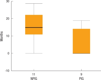
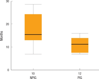
From September 2004 to January 2008, 266 patients who were treated for colorectal cancer at Department of Surgery, Hanyang University College of Medicine, were divided into two groups: one that underwent preoperative and postoperative periodical chest CT (periodical inspection group, PIG; May 2006 to January 2008, 135 patients) and one that did not undergo periodical chest CT (non-periodical inspection group, NPIG; September 2004 to April 2006, 131 patients) for comparison.
The overall lung metastasis diagnosis rates did not manifest any significant difference. The times to diagnose lung metastasis patients were 6.3 months and 15.7 months for the PIG and the NPIG, respectively (P = 0.022). The size of the metastatic lung nodule was smaller in the PIG than in the NPIG (< 1 cm in 9/9 patients vs. < 1 cm in 6/9 patients in the PIG and the NPIG, respectively; P = 0.02). A solitary lung metastasis was more frequently found in the PIG (5/9 patients) than in the NPIG (1/11 patients) (P = 0.024). During the follow-up period, 100% (2/2 patients) and 60% (3/5 patients) of the patients in the PIG and the NPIG, respectively, with stage III cancer underwent a lung metastasectomy (P = 0.002).
Chest CT enables early diagnosis with a smaller size and a lower number of lung metastases in patients with colorectal cancer. Moreover, pulmonary the rate of the pulmonary resection for selected patients may be improved. However, the contribution of chest CT to increasing the survival rate must be investigated in a prospective randomized study.
本研究旨在确定胸部计算机断层扫描(CT)在结直肠癌肺转移诊断中的实用价值。
2004年9月至2008年1月,在汉阳大学医学院外科接受结直肠癌治疗的266例患者被分为两组:一组在术前和术后进行定期胸部CT检查(定期检查组,PIG;2006年5月至2008年1月,135例患者),另一组未进行定期胸部CT检查(非定期检查组,NPIG;2004年9月至2006年4月,131例患者)进行比较。
总体肺转移诊断率无显著差异。PIG组和NPIG组诊断肺转移患者的时间分别为6.3个月和15.7个月(P = 0.022)。PIG组转移肺结节的大小小于NPIG组(PIG组9例患者中有9例<1 cm,NPIG组9例患者中有6例<1 cm;P = 0.02)。PIG组(5/9例患者)比NPIG组(1/11例患者)更常发现孤立性肺转移(P = 0.024)。在随访期间,PIG组和NPIG组中分别有100%(2/2例患者)和60%(3/5例患者)的III期癌症患者接受了肺转移瘤切除术(P = 0.002)。
胸部CT能够对结直肠癌患者进行早期诊断,且肺转移灶数量较少、体积较小。此外,对于选定患者的肺切除率可能会提高。然而,胸部CT对提高生存率的贡献必须在前瞻性随机研究中进行调查。