Hospital for Children and Adolescents, Helsinki University Central Hospital, Tukholmankatu 2C, PO Box 705, 00029 Helsinki, Finland.
Osteoporos Int. 2011 Mar;22(3):883-91. doi: 10.1007/s00198-010-1499-4. Epub 2010 Dec 10.
In this prospective study, 87 children were followed up from birth to 14 months with data on maternal vitamin D status during the pregnancy. Postnatal vitamin D supplementation improved vitamin D status but only partly eliminated the differences in bone variables induced by maternal vitamin D status during the fetal period.
Intrauterine nutritional deficits may have permanent consequences despite improved nutritional status postnatally. We evaluated the role of prenatal and postnatal vitamin D status on bone parameters in early infancy.
Eighty-seven children were followed from birth to 14 months. Background data were collected with a questionnaire and a 3-day food record. At 14 months bone variables were measured with peripheral computed tomography (pQCT) from the left tibia. Serum 25-OHD and bone turnover markers were determined. Findings were compared with maternal vitamin D status during pregnancy.
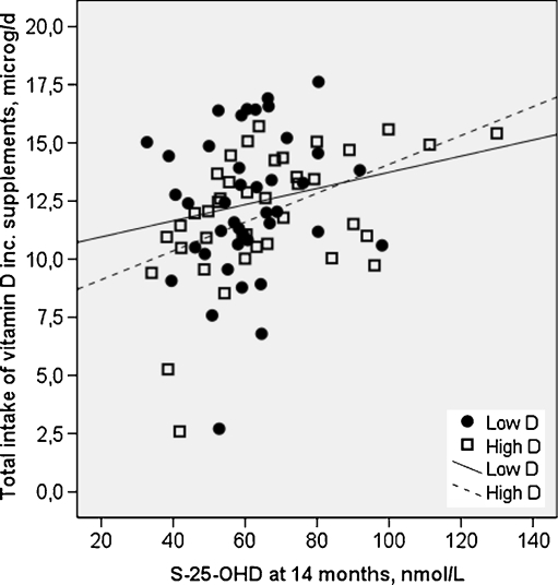
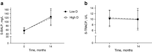
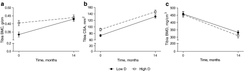
The children were divided into two groups based on vitamin D status during pregnancy. Despite discrepant S-25-OHD at baseline (median 36.3 vs. 52.5 nmol/l, p < 0.001), the values at 14 months were similar (63 vs. 66 nmol/l, p = 0.58) in Low D and High D. Serum 25-OHD increased more in Low D (p < 0.001) despite similar total intake of vitamin D (mean 12.3 μg/day). In Low D, tibial bone mineral content (BMC) was lower at birth but BMC gain was greater (multivariate analysis of variance [MANOVA]; p = 0.032) resulting in similar BMC at 14 months in the two groups. In High D, tibial total bone cross-sectional area was higher at baseline; the difference persisted at 14 months (MANOVA; p = 0.068). Bone mineral density (BMD) and ΔBMD were similar in the two groups.
Postnatal vitamin D supplementation improved vitamin D status but only partly eliminated the differences in bone variables induced by maternal vitamin D status during the fetal period. Further attention should be paid to improving vitamin D status during pregnancy.
在这项前瞻性研究中,87 名儿童从出生到 14 个月接受了随访,数据涉及母亲怀孕期间的维生素 D 状况。产后维生素 D 补充改善了维生素 D 状况,但仅部分消除了胎儿期母体维生素 D 状况对骨骼变量的影响。
尽管出生后营养状况得到改善,但宫内营养不足仍可能产生持久影响。我们评估了产前和产后维生素 D 状况对婴儿早期骨骼参数的作用。
87 名儿童从出生到 14 个月接受了随访。通过问卷调查和 3 天食物记录收集背景数据。在 14 个月时,使用外周计算机断层扫描(pQCT)从左胫骨测量骨骼变量。测定血清 25-羟维生素 D 和骨转换标志物。结果与妊娠期间的母体维生素 D 状况进行比较。
根据妊娠期间的维生素 D 状况,将儿童分为两组。尽管基线时血清 25-羟维生素 D 值存在差异(中位数 36.3 与 52.5 nmol/L,p<0.001),但 14 个月时两组的血清 25-羟维生素 D 值相似(63 与 66 nmol/L,p=0.58)。尽管维生素 D 总摄入量相同(平均 12.3 μg/天),但在低维生素 D 组中血清 25-羟维生素 D 增加更多(p<0.001)。在低维生素 D 组中,出生时胫骨骨矿物质含量(BMC)较低,但 BMC 增加更大(多变量方差分析[MANOVA];p=0.032),导致两组 14 个月时 BMC 相似。在高维生素 D 组中,基线时胫骨总骨横截面积较高;差异在 14 个月时仍存在(MANOVA;p=0.068)。两组的骨密度(BMD)和 ΔBMD 相似。
产后维生素 D 补充改善了维生素 D 状况,但仅部分消除了胎儿期母体维生素 D 状况对骨骼变量的影响。应进一步关注改善妊娠期间的维生素 D 状况。