Department of Radiation Oncology, Klinikum rechts der Isar, Technische Universität München, Munich, Germany.
PLoS One. 2010 Dec 16;5(12):e15339. doi: 10.1371/journal.pone.0015339.
Apart from the platelet/endothelial cell adhesion molecule 1 (PECAM-1, CD31), endoglin (CD105) and a positive factor VIII-related antigen staining, human primary and immortalized macro- and microvascular endothelial cells (ECs) differ in their cell surface expression of activating and inhibitory ligands for natural killer (NK) cells. Here we comparatively study the effects of irradiation on the phenotype of ECs and their interaction with resting and activated NK cells.
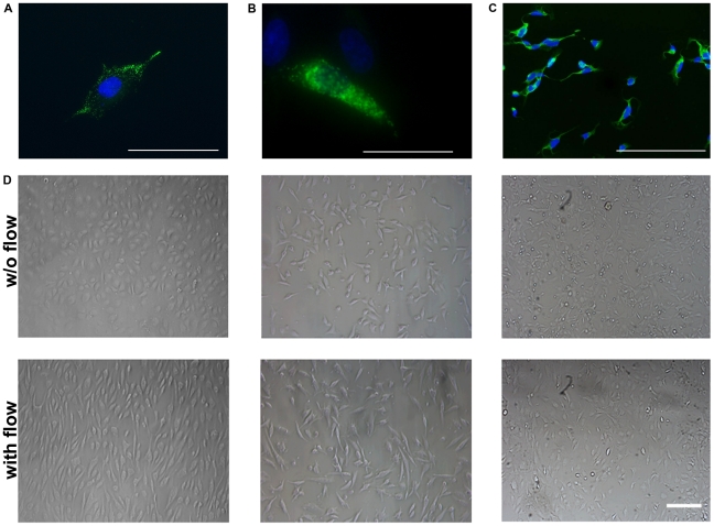
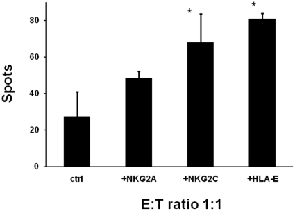
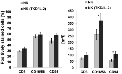
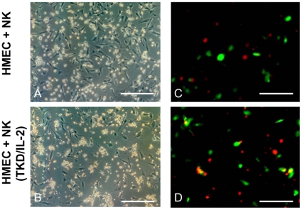
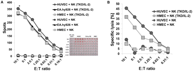
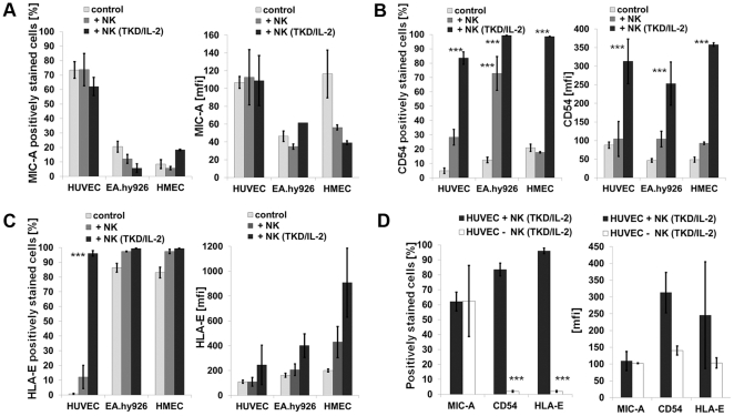
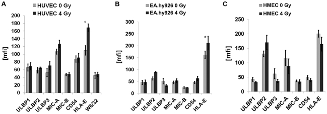
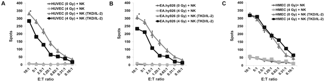
METHODOLOGY/PRINCIPAL FINDINGS: Primary macrovascular human umbilical vein endothelial cells (HUVECs) only express UL16 binding protein 2 (ULBP2) and the major histocompatibility complex (MHC) class I chain-related protein MIC-A (MIC-A) as activating signals for NK cells, whereas the corresponding immortalized EA.hy926 EC cell line additionally present ULBP3, membrane heat shock protein 70 (Hsp70), intercellular adhesion molecule ICAM-1 (CD54) and HLA-E. Apart from MIC-B, the immortalized human microvascular endothelial cell line HMEC, resembles the phenotype of EA.hy926. Surprisingly, primary HUVECs are more sensitive to Hsp70 peptide (TKD) plus IL-2 (TKD/IL-2)-activated NK cells than their immortalized EC counterpatrs. This finding is most likely due to the absence of the inhibitory ligand HLA-E, since the activating ligands are shared among the ECs. The co-culture of HUVECs with activated NK cells induces ICAM-1 (CD54) and HLA-E expression on the former which drops to the initial low levels (below 5%) when NK cells are removed. Sublethal irradiation of HUVECs induces similar but less pronounced effects on HUVECs. Along with these findings, irradiation also induces HLA-E expression on macrovascular ECs and this correlates with an increased resistance to killing by activated NK cells. Irradiation had no effect on HLA-E expression on microvascular ECs and the sensitivity of these cells to NK cells remained unaffected.
CONCLUSION/SIGNIFICANCE: These data emphasize that an irradiation-induced, transient up-regulation of HLA-E on macrovascular ECs might confer protection against NK cell-mediated vascular injury.
除血小板/内皮细胞黏附分子 1(PECAM-1,CD31)、内皮糖蛋白(CD105)和 VIII 因子相关抗原阳性染色外,人原代和永生化的大、微血管内皮细胞(EC)在其表面表达的自然杀伤(NK)细胞激活和抑制配体方面存在差异。在此,我们比较研究了辐照对 EC 表型及其与静息和激活 NK 细胞相互作用的影响。
方法/主要发现:原代大血管人脐静脉内皮细胞(HUVEC)仅表达 UL16 结合蛋白 2(ULBP2)和主要组织相容性复合体(MHC)I 类链相关蛋白 MIC-A(MIC-A)作为 NK 细胞的激活信号,而相应的永生化 EA.hy926 EC 细胞系另外还表达 ULBP3、膜热休克蛋白 70(Hsp70)、细胞间黏附分子 ICAM-1(CD54)和 HLA-E。除 MIC-B 外,永生化人微血管内皮细胞系 HMEC 与 EA.hy926 的表型相似。令人惊讶的是,与永生化 EC 对照物相比,原代 HUVEC 对 HSP70 肽(TKD)加 IL-2(TKD/IL-2)激活的 NK 细胞更为敏感。这一发现很可能是由于缺乏抑制性配体 HLA-E,因为激活配体在 EC 之间共享。与激活的 NK 细胞共培养诱导 HUVEC 表达 ICAM-1(CD54)和 HLA-E,当 NK 细胞被去除时,前者的表达降至初始低水平(低于 5%)。亚致死剂量的辐照诱导 HUVEC 产生类似但不太明显的影响。随着这些发现,辐照还诱导大血管 EC 表达 HLA-E,这与对激活的 NK 细胞杀伤的抵抗力增加相关。辐照对微血管 EC 上 HLA-E 的表达没有影响,这些细胞对 NK 细胞的敏感性保持不变。
结论/意义:这些数据强调,血管内皮细胞上 HLA-E 的辐照诱导、短暂上调可能为 NK 细胞介导的血管损伤提供保护。