Division of Cardiology, Research Center of Cardiovascular Biology, University of Genova, Genova, Italy.
PLoS One. 2010 Dec 20;5(12):e15583. doi: 10.1371/journal.pone.0015583.
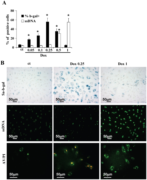
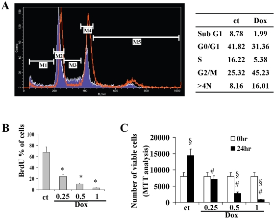
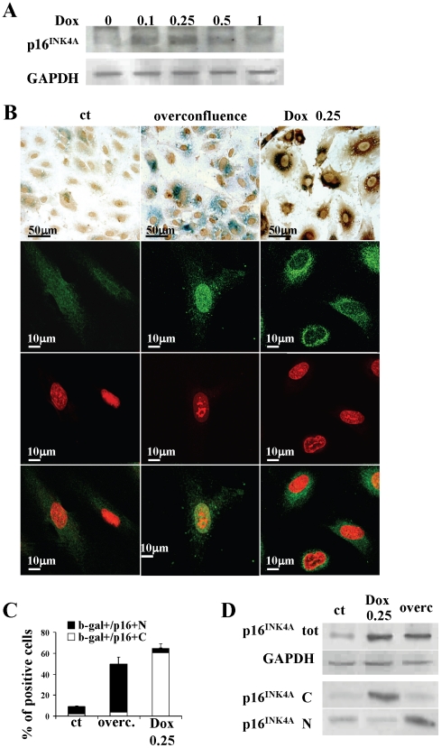
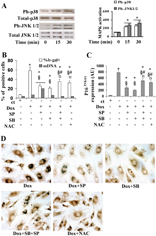
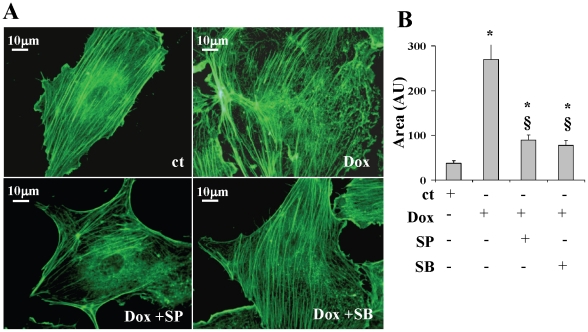
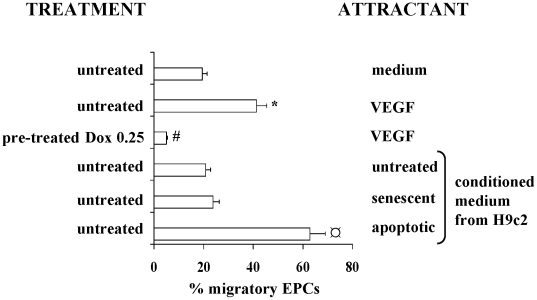
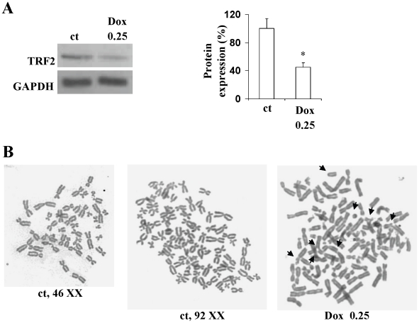
Patients treated with low-dose anthracyclines often show late onset cardiotoxicity. Recent studies suggest that this form of cardiotoxicity is the result of a progenitor cell disease. In this study we demonstrate that Cord Blood Endothelial Progenitor Cells (EPCs) exposed to low, sub-apoptotic doses of doxorubicin show a senescence phenotype characterized by increased SA-b-gal activity, decreased TRF2 and chromosomal abnormalities, enlarged cell shape, and disarrangement of F-actin stress fibers accompanied by impaired migratory ability. P16( INK4A) localizes in the cytoplasm of doxorubicin-induced senescent EPCs and not in the nucleus as is the case in EPCs rendered senescent by different stimuli. This localization together with the presence of an arrest in G2, and not at the G1 phase boundary, which is what usually occurs in response to the cell cycle regulatory activity of p16(INK4A), suggests that doxorubicin-induced p16( INK4A) does not regulate the cell cycle, even though its increase is closely associated with senescence. The effects of doxorubicin are the result of the activation of MAPKs p38 and JNK which act antagonistically. JNK attenuates the senescence, p16( INK4A) expression and cytoskeleton remodeling that are induced by activated p38. We also found that conditioned medium from doxorubicin-induced senescent cardiomyocytes does not attract untreated EPCs, unlike conditioned medium from apoptotic cardiomyocytes which has a strong chemoattractant capacity. In conclusion, this study provides a better understanding of the senescence of doxorubicin-treated EPCs, which may be helpful in preventing and treating late onset cardiotoxicity.
接受低剂量蒽环类药物治疗的患者常出现迟发性心脏毒性。最近的研究表明,这种形式的心脏毒性是前体细胞疾病的结果。在这项研究中,我们证明暴露于低剂量亚凋亡剂量阿霉素的脐血内皮祖细胞(EPC)表现出衰老表型,其特征为 SA-b-gal 活性增加、TRF2 和染色体异常减少、细胞形状增大以及 F-肌动蛋白应力纤维排列紊乱,同时迁移能力受损。P16(INK4A)定位于阿霉素诱导的衰老 EPC 的细胞质中,而不是细胞核中,如不同刺激诱导的 EPC 衰老时那样。这种定位以及 G2 期停滞而不是通常发生在 p16(INK4A)对细胞周期调节活性的 G1 期边界的停滞,表明阿霉素诱导的 p16(INK4A)不调节细胞周期,尽管其增加与衰老密切相关。阿霉素的作用是 MAPKs p38 和 JNK 激活的结果,它们拮抗作用。JNK 减弱了由激活的 p38 诱导的衰老、p16(INK4A)表达和细胞骨架重塑。我们还发现,与凋亡性心肌细胞的条件培养基具有很强的趋化能力不同,来自阿霉素诱导的衰老心肌细胞的条件培养基不会吸引未处理的 EPC。总之,这项研究提供了对阿霉素处理的 EPC 衰老的更好理解,这可能有助于预防和治疗迟发性心脏毒性。