Department of Biology, University of Virginia, Charlottesville, Virginia, United States of America.
PLoS One. 2010 Dec 20;5(12):e15599. doi: 10.1371/journal.pone.0015599.
Although an endogenous circadian clock located in the retinal photoreceptor layer governs various physiological events including melatonin rhythms in Xenopus laevis, it remains unknown which of the photoreceptors, rod and/or cone, is responsible for the circadian regulation of melatonin release.
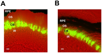
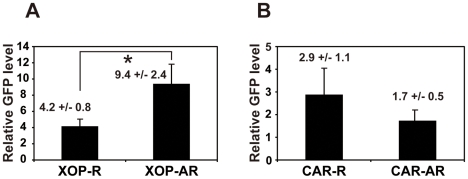
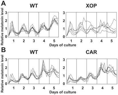
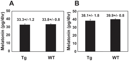
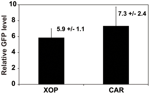
METHODOLOGY/PRINCIPAL FINDINGS: We selectively disrupted circadian clock function in either the rod or cone photoreceptor cells by generating transgenic Xenopus tadpoles expressing a dominant-negative CLOCK (XCLΔQ) under the control of a rod or cone-specific promoter. Eyecup culture and continuous melatonin measurement revealed that circadian rhythms of melatonin release were abolished in a majority of the rod-specific XCLΔQ transgenic tadpoles, although the percentage of arrhythmia was lower than that of transgenic tadpole eyes expressing XCLΔQ in both rods and cones. In contrast, whereas a higher percentage of arrhythmia was observed in the eyes of the cone-specific XCLΔQ transgenic tadpoles compare to wild-type counterparts, the rate was significantly lower than in rod-specific transgenics. The levels of the transgene expression were comparable between these two different types of transgenics. In addition, the average overall melatonin levels were not changed in the arrhythmic eyes, suggesting that CLOCK does not affect absolute levels of melatonin, only its temporal expression pattern.
CONCLUSIONS/SIGNIFICANCE: These results suggest that although the Xenopus retina is made up of approximately equal numbers of rods and cones, the circadian clocks in the rod cells play a dominant role in driving circadian melatonin rhythmicity in the Xenopus retina, although some contribution of the clock in cone cells cannot be excluded.
虽然位于视网膜光感受器层的内源性生物钟控制着各种生理事件,包括非洲爪蟾的褪黑素节律,但尚不清楚是杆状细胞和/或锥状细胞中的哪一种负责褪黑素释放的生物钟调节。
方法/主要发现:我们通过在杆状细胞或锥状细胞光感受器细胞中表达显性负性 CLOCK(XCLΔQ),生成转基因非洲爪蟾蝌蚪,选择性地破坏生物钟功能,该基因由杆状细胞或锥状细胞特异性启动子控制。眼杯培养和连续褪黑素测量显示,大多数杆状细胞特异性 XCLΔQ 转基因蝌蚪的褪黑素释放节律被消除,尽管节律紊乱的百分比低于同时在杆状细胞和锥状细胞中表达 XCLΔQ 的转基因蝌蚪眼。相比之下,尽管在锥状细胞特异性 XCLΔQ 转基因蝌蚪眼中观察到的节律紊乱百分比高于野生型对照,但该比率明显低于杆状细胞特异性转基因。两种不同类型的转基因之间,转基因表达水平相当。此外,在这些节律紊乱的眼中,平均整体褪黑素水平没有变化,表明 CLOCK 不影响褪黑素的绝对水平,仅影响其时间表达模式。
结论/意义:这些结果表明,尽管非洲爪蟾的视网膜由大约等量的杆状细胞和锥状细胞组成,但杆状细胞中的生物钟在驱动非洲爪蟾视网膜中的生物钟褪黑素节律方面起着主导作用,尽管不能排除锥状细胞时钟的一些贡献。