• 文献检索
  • 文档翻译
  • 深度研究
  • 学术资讯
  • Suppr Zotero 插件Zotero 插件
  • 邀请有礼
  • 套餐&价格
  • 历史记录
应用&插件
Suppr Zotero 插件Zotero 插件浏览器插件Mac 客户端Windows 客户端微信小程序
定价
高级版会员购买积分包购买API积分包
服务
文献检索文档翻译深度研究API 文档MCP 服务
关于我们
关于 Suppr公司介绍联系我们用户协议隐私条款
关注我们

Suppr 超能文献

核心技术专利:CN118964589B侵权必究
粤ICP备2023148730 号-1Suppr @ 2026

文献检索

告别复杂PubMed语法,用中文像聊天一样搜索,搜遍4000万医学文献。AI智能推荐,让科研检索更轻松。

立即免费搜索

文件翻译

保留排版,准确专业,支持PDF/Word/PPT等文件格式,支持 12+语言互译。

免费翻译文档

深度研究

AI帮你快速写综述,25分钟生成高质量综述,智能提取关键信息,辅助科研写作。

立即免费体验

用于自适应钴断层放射治疗的磁共振成像:一项提议。

Magnetic resonance imaging for adaptive cobalt tomotherapy: A proposal.

作者信息

Kron Tomas, Eyles David, Schreiner L John, Battista Jerry

机构信息

Peter MacCallum Cancer Centre, Melbourne, Australia.

出版信息

J Med Phys. 2006 Oct;31(4):242-54. doi: 10.4103/0971-6203.29194.

DOI:10.4103/0971-6203.29194
PMID:21206640
原文链接:https://pmc.ncbi.nlm.nih.gov/articles/PMC3004099/
Abstract

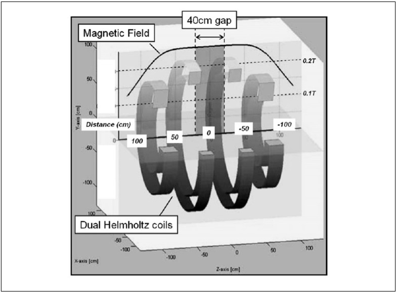
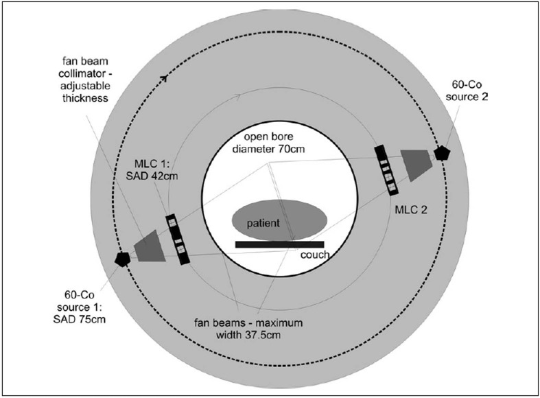
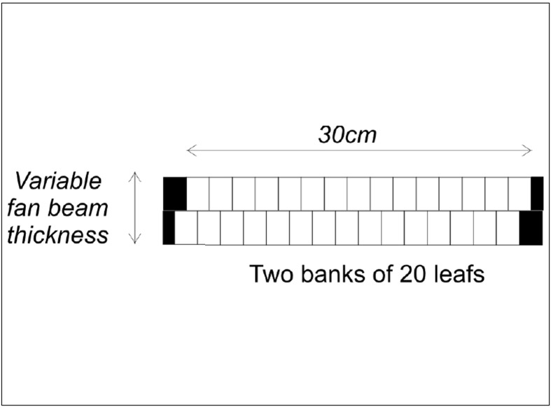
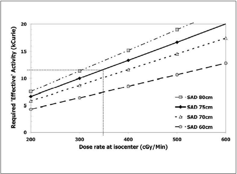
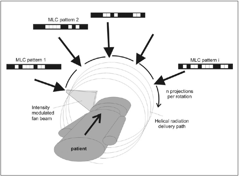
Magnetic resonance imaging (MRI) provides excellent soft tissue contrast for oncology applications. We propose to combine a MRI scanner with a helical tomotherapy (HT) system to enable daily target imaging for improved conformal radiation dose delivery to a patient. HT uses an intensity-modulated fan-beam that revolves around a patient, while the patient slowly advances through the plane of rotation, yielding a helical beam trajectory. Since the use of a linear accelerator to produce radiation may be incompatible with the pulsed radiofrequency and the high and pulsed magnetic fields required for MRI, it is proposed that a radioactive Cobalt-60 ((60)Co) source be used instead to provide the radiation. An open low field (0.25 T) MRI system is proposed where the tomotherapy ring gantry is located between two sets of Helmholtz coils that can generate a sufficiently homogenous main magnetic field.It is shown that the two major challenges with the design, namely acceptable radiation dose rate (and therefore treatment duration) and moving parts in strong magnetic field, can be addressed. The high dose rate desired for helical tomotherapy delivery can be achieved using two radiation sources of 220TBq (6000Ci) each on a ring gantry with a source to axis-of-rotation distance of 75 cm. In addition to this, a dual row multi-leaf collimator (MLC) system with 15 mm leaf width at isocentre and relatively large fan beam widths between 15 and 30 mm per row shall be employed. In this configuration, the unit would be well-suited for most pelvic radiotherapy applications where the soft tissue contrast of MRI will be particularly beneficial. Non-magnetic MRI compatible materials must be used for the rotating gantry. Tungsten, which is non-magnetic, can be used for primary collimation of the fan-beam as well as for the MLC, which allows intensity modulated radiation delivery. We propose to employ a low magnetic Cobalt compound, sycoporite (CoS) for the Cobalt source material itself.Rotational delivery is less susceptible to problems related to the use of a low energy megavoltage photon source while the helical delivery reduces the negative impact of the relatively large penumbra inherent in the use of Cobalt sources for radiotherapy. On the other hand, the use of a (60)Co source ensures constant dose rate with gantry rotation and makes dose calculation in a magnetic field as easy as the range of secondary electrons is limited.The MR-integrated Cobalt tomotherapy unit, dubbed 'MiCoTo,' uses two independent physical principles for image acquisition and treatment delivery. It would offer excellent target definition and will allow following target motion during treatment using fast imaging techniques thus providing the best possible input for adaptive radiotherapy. As an additional bonus, quality assurance of the radiation delivery can be performed in situ using radiation sensitive gels imaged by MRI.

https://cdn.ncbi.nlm.nih.gov/pmc/blobs/6915/3004099/48170c74f314/JMP-31-242-g008.jpg
https://cdn.ncbi.nlm.nih.gov/pmc/blobs/6915/3004099/9472bc38ae7e/JMP-31-242-g001.jpg
https://cdn.ncbi.nlm.nih.gov/pmc/blobs/6915/3004099/57e1d11a3e7c/JMP-31-242-g002.jpg
https://cdn.ncbi.nlm.nih.gov/pmc/blobs/6915/3004099/4aae166c8898/JMP-31-242-g003.jpg
https://cdn.ncbi.nlm.nih.gov/pmc/blobs/6915/3004099/17f7fa7289bf/JMP-31-242-g004.jpg
https://cdn.ncbi.nlm.nih.gov/pmc/blobs/6915/3004099/e0979d210971/JMP-31-242-g005.jpg
https://cdn.ncbi.nlm.nih.gov/pmc/blobs/6915/3004099/bf652ad88c10/JMP-31-242-g006.jpg
https://cdn.ncbi.nlm.nih.gov/pmc/blobs/6915/3004099/eec0ae74c8e2/JMP-31-242-g007.jpg
https://cdn.ncbi.nlm.nih.gov/pmc/blobs/6915/3004099/48170c74f314/JMP-31-242-g008.jpg
摘要

磁共振成像(MRI)在肿瘤学应用中能提供出色的软组织对比度。我们提议将MRI扫描仪与螺旋断层放射治疗(HT)系统相结合,以便每天对靶区进行成像,从而更精确地向患者输送适形放射剂量。HT使用强度调制扇形束,围绕患者旋转,同时患者缓慢通过旋转平面,形成螺旋形束轨迹。由于使用直线加速器产生辐射可能与MRI所需的脉冲射频以及高脉冲磁场不兼容,因此建议改用放射性钴 - 60(⁶⁰Co)源来提供辐射。提议采用开放式低场(0.25T)MRI系统,其中断层放射治疗环形机架位于两组亥姆霍兹线圈之间,这两组线圈可产生足够均匀的主磁场。结果表明,该设计面临的两个主要挑战,即可接受的放射剂量率(以及由此决定的治疗持续时间)和强磁场中的运动部件,是可以解决的。使用两个220TBq(6000Ci)的放射源,放置在环形机架上,源到旋转轴的距离为75cm,可实现螺旋断层放射治疗所需的高剂量率。除此之外,应采用在等中心处叶片宽度为15mm且每行扇束宽度在15至30mm之间相对较大的双排多叶准直器(MLC)系统。在这种配置下,该装置将非常适合大多数盆腔放射治疗应用,其中MRI的软组织对比度将特别有益。旋转机架必须使用与MRI兼容的非磁性材料。非磁性的钨可用于扇形束的初级准直以及MLC,从而实现强度调制放射输送。我们提议使用低磁性的钴化合物西科波石(CoS)作为钴源材料本身。旋转式输送对与使用低能兆伏光子源相关的问题不太敏感,而螺旋式输送减少了钴源用于放射治疗时固有的相对较大半值层的负面影响。另一方面,使用⁶⁰Co源可确保随着机架旋转剂量率恒定,并且由于二次电子的射程有限,使得在磁场中进行剂量计算变得容易。集成MRI的钴断层放射治疗装置,称为“MiCoTo”,使用两种独立的物理原理进行图像采集和治疗输送。它将提供出色的靶区定义,并允许在治疗期间使用快速成像技术跟踪靶区运动,从而为自适应放射治疗提供尽可能好的输入。另外,使用MRI成像的辐射敏感凝胶可在原位对放射输送进行质量保证。

相似文献

1
Magnetic resonance imaging for adaptive cobalt tomotherapy: A proposal.用于自适应钴断层放射治疗的磁共振成像:一项提议。
J Med Phys. 2006 Oct;31(4):242-54. doi: 10.4103/0971-6203.29194.
2
Multibeam tomotherapy: a new treatment unit devised for multileaf collimation, intensity-modulated radiation therapy.多叶准直器调强放射治疗用多束断层放射治疗设备:一种新的治疗装置
Med Phys. 2007 Oct;34(10):3926-42. doi: 10.1118/1.2779129.
3
Tomotherapy as a tool in image-guided radiation therapy (IGRT): theoretical and technological aspects.螺旋断层放疗作为图像引导放射治疗(IGRT)中的一种工具:理论与技术层面
Biomed Imaging Interv J. 2007 Jan;3(1):e16. doi: 10.2349/biij.3.1.e16. Epub 2007 Jan 1.
4
Monte Carlo calculation of helical tomotherapy dose delivery.螺旋断层放射治疗剂量输出的蒙特卡罗计算
Med Phys. 2008 Aug;35(8):3491-500. doi: 10.1118/1.2948409.
5
Investigation of an efficient source design for Cobalt-60-based tomotherapy using EGSnrc Monte Carlo simulations.使用EGSnrc蒙特卡罗模拟对基于钴-60的断层放射治疗的高效源设计进行研究。
Phys Med Biol. 2008 Feb 7;53(3):575-92. doi: 10.1088/0031-9155/53/3/005. Epub 2008 Jan 10.
6
Results of a two-year quality control program for a helical tomotherapy unit.螺旋断层放射治疗设备的两年质量控制计划结果
Radiother Oncol. 2008 Feb;86(2):231-41. doi: 10.1016/j.radonc.2007.11.005. Epub 2007 Dec 3.
7
Tomotherapy.断层放射治疗
Semin Radiat Oncol. 1999 Jan;9(1):108-17. doi: 10.1016/s1053-4296(99)80058-7.
8
Filmless methods for quality assurance of Tomotherapy using ArcCHECK.使用ArcCHECK对螺旋断层放疗进行质量保证的无胶片方法。
Med Phys. 2017 Jan;44(1):7-16. doi: 10.1002/mp.12009. Epub 2017 Jan 3.
9
Commissioning experience and quality assurance of helical tomotherapy machines.螺旋断层放射治疗机的调试经验与质量保证
J Med Phys. 2009 Oct;34(4):194-9. doi: 10.4103/0971-6203.56078.
10
A topographic leaf-sequencing algorithm for delivering intensity modulated radiation therapy.一种用于实施调强放射治疗的断层叶片排序算法。
Med Phys. 2006 Aug;33(8):2751-6. doi: 10.1118/1.2216876.

引用本文的文献

1
Erratum: Magnetic Resonance Imaging for Adaptive Cobalt Tomotherapy: A Proposal.勘误:用于自适应钴-60治疗的磁共振成像:一项提议。
J Med Phys. 2018 Apr-Jun;43(2):146. doi: 10.4103/0971-6203.234536.
2
Prospective feasibility analysis of a novel off-line approach for MR-guided radiotherapy.新型离线 MR 引导放疗方法的前瞻性可行性分析。
Strahlenther Onkol. 2018 May;194(5):425-434. doi: 10.1007/s00066-017-1258-y. Epub 2018 Jan 18.
3
The future of image-guided radiotherapy will be MR guided.图像引导放射治疗的未来将是磁共振引导的。

本文引用的文献

1
The use of MRI in planning radiotherapy for gynaecological tumours.磁共振成像在妇科肿瘤放射治疗计划中的应用。
Cancer Imaging. 2006 Jun 22;6(1):100-6. doi: 10.1102/1470-7330.2006.0016.
2
Efficient on-line setup correction strategies using plan-intent functions.使用计划意图函数的高效在线设置校正策略。
Med Phys. 2006 May;33(5):1388-97. doi: 10.1118/1.2192580.
3
A comparison of CT- and ultrasound-based imaging to localize the prostate for external beam radiotherapy.基于CT和超声成像在体外束放疗中对前列腺进行定位的比较。
Br J Radiol. 2017 May;90(1073):20160667. doi: 10.1259/bjr.20160667. Epub 2017 Mar 29.
4
Monte Carlo simulations of patient dose perturbations in rotational-type radiotherapy due to a transverse magnetic field: a tomotherapy investigation.旋转式放射治疗中横向磁场引起的患者剂量扰动的蒙特卡罗模拟:一项断层放射治疗研究。
Med Phys. 2015 Feb;42(2):715-25. doi: 10.1118/1.4905168.
5
Prospective observer and software-based assessment of magnetic resonance imaging quality in head and neck cancer: Should standard positioning and immobilization be required for radiation therapy applications?头颈部癌磁共振成像质量的前瞻性观察者及基于软件的评估:放射治疗应用是否需要标准定位和固定?
Pract Radiat Oncol. 2015 Jul-Aug;5(4):e299-308. doi: 10.1016/j.prro.2014.11.003. Epub 2014 Dec 17.
6
Registration accuracy for MR images of the prostate using a subvolume based registration protocol.基于子体积的配准协议对前列腺磁共振图像的配准精度。
Radiat Oncol. 2011 Jun 16;6:73. doi: 10.1186/1748-717X-6-73.
7
The role of Cobalt-60 in modern radiation therapy: Dose delivery and image guidance.钴-60在现代放射治疗中的作用:剂量输送与图像引导。
J Med Phys. 2009 Jul;34(3):133-6. doi: 10.4103/0971-6203.54846.
8
Reduction of margins in external beam radiotherapy.外照射放疗中 margins 的缩减。
J Med Phys. 2008 Apr;33(2):41-2. doi: 10.4103/0971-6203.41190.
9
Assessment of dose reconstruction errors in image-guided radiation therapy.图像引导放射治疗中剂量重建误差的评估。
Phys Med Biol. 2008 Feb 7;53(3):719-36. doi: 10.1088/0031-9155/53/3/013. Epub 2008 Jan 11.
Int J Radiat Oncol Biol Phys. 2006 Jul 1;65(3):678-87. doi: 10.1016/j.ijrobp.2006.01.022.
4
Evaluation of possible prostate displacement induced by pressure applied during transabdominal ultrasound image acquisition.经腹超声图像采集过程中施加压力导致前列腺可能移位的评估。
Strahlenther Onkol. 2006 Apr;182(4):240-6. doi: 10.1007/s00066-006-1513-0.
5
Improved target volume definition for fractionated stereotactic radiotherapy in patients with intracranial meningiomas by correlation of CT, MRI, and [68Ga]-DOTATOC-PET.通过CT、MRI和[68Ga]-DOTATOC-PET的相关性改进颅内脑膜瘤患者分次立体定向放射治疗的靶区定义
Int J Radiat Oncol Biol Phys. 2006 May 1;65(1):222-7. doi: 10.1016/j.ijrobp.2005.12.006. Epub 2006 Feb 20.
6
Integrated PET/CT: current applications and future directions.一体化正电子发射断层显像/计算机断层扫描(PET/CT):当前应用及未来发展方向
Radiology. 2006 Feb;238(2):405-22. doi: 10.1148/radiol.2382041977.
7
A method for deconvolution of integrated electronic portal images to obtain incident fluence for dose reconstruction.一种用于对集成电子射野影像进行去卷积以获取用于剂量重建的入射注量的方法。
J Appl Clin Med Phys. 2005 Fall;6(4):22-39. doi: 10.1120/jacmp.v6i4.2104. Epub 2005 Nov 21.
8
From IMRT to IGRT: frontierland or neverland?从调强放疗到图像引导放疗:前沿领域还是虚幻之地?
Radiother Oncol. 2006 Feb;78(2):119-22. doi: 10.1016/j.radonc.2005.12.005. Epub 2006 Jan 18.
9
Towards truly integrated hardware fusion with PET/CT.迈向与PET/CT真正集成的硬件融合。
Nuklearmedizin. 2005;44 Suppl 1:S5-12.
10
Radiotherapeutic techniques for prostate cancer, dose escalation and brachytherapy.前列腺癌的放射治疗技术、剂量递增及近距离放射治疗
Clin Oncol (R Coll Radiol). 2005 Oct;17(7):560-71. doi: 10.1016/j.clon.2005.07.006.