Laboratori d'Enginyeria de Proteïnes, Departament de Biologia, Facultat de Ciències, Universitat de Girona, Campus de Montilivi s/n E-17071 Girona, Spain.
BMC Cancer. 2011 Jan 11;11:9. doi: 10.1186/1471-2407-11-9.
Ribonucleases are promising agents for use in anticancer therapy. Among the different ribonucleases described to be cytotoxic, a paradigmatic example is onconase which manifests cytotoxic and cytostatic effects, presents synergism with several kinds of anticancer drugs and is currently in phase II/III of its clinical trial as an anticancer drug against different types of cancer. The mechanism of cytotoxicity of PE5, a variant of human pancreatic ribonuclease carrying a nuclear localization signal, has been investigated and compared to that of onconase.
Cytotoxicity was measured by the MTT method and by the tripan blue exclusion assay. Apoptosis was assessed by flow cytometry, caspase enzymatic detection and confocal microscopy. Cell cycle phase analysis was performed by flow cytometry. The expression of different proteins was analyzed by western blot.
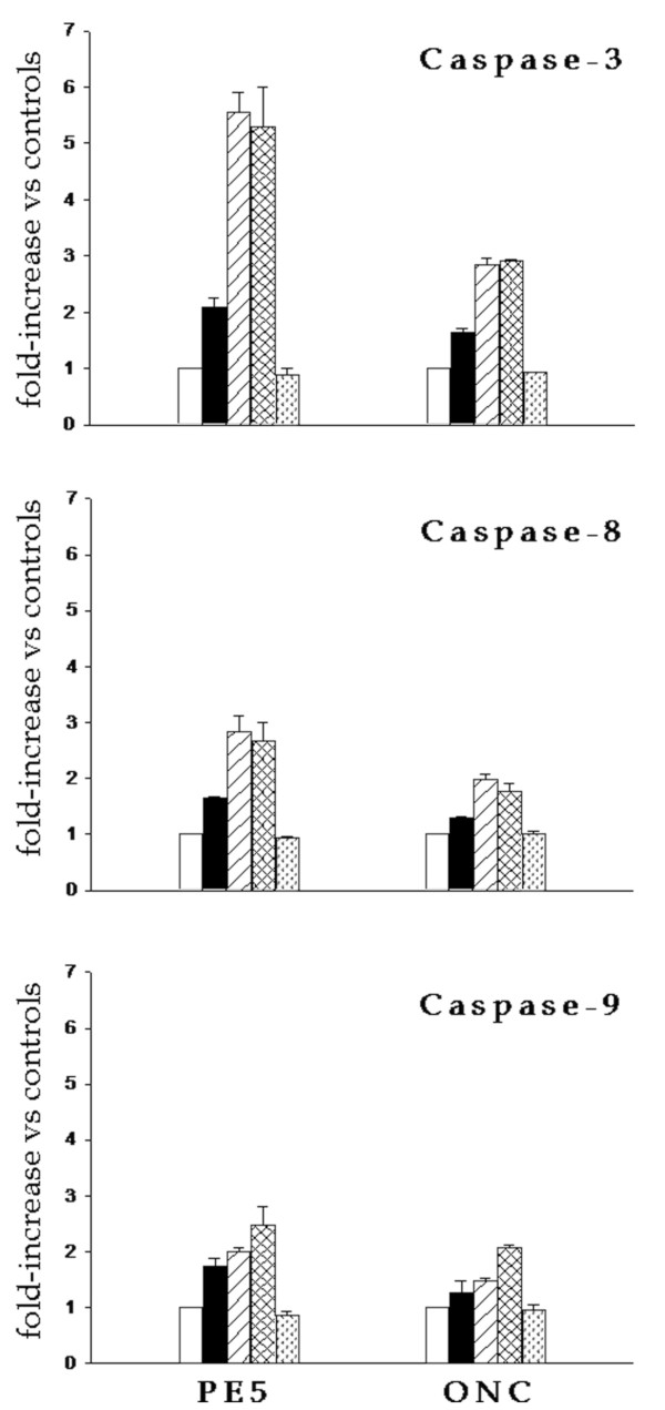
We show that the cytotoxicity of PE5 is produced through apoptosis, that it does not require the proapoptotic activity of p53 and is not prevented by the multiple drug resistance phenotype. We also show that PE5 and onconase induce cell death at the same extent although the latter is also able to arrest the cell growth. We have compared the cytotoxic effects of both ribonucleases in the NCI/ADR-RES cell line by measuring their effects on the cell cycle, on the activation of different caspases and on the expression of different apoptosis- and cell cycle-related proteins. PE5 increases the number of cells in S and G2/M cell cycle phases, which is accompanied by the increased expression of cyclin E and p21WAF1/CIP1 together with the underphosphorylation of p46 forms of JNK. Citotoxicity of onconase in this cell line does not alter the cell cycle phase distribution and it is accompanied by a decreased expression of XIAP CONCLUSIONS: We conclude that PE5 kills the cells through apoptosis associated with the p21WAF1/CIP1 induction and the inactivation of JNK. This mechanism is significantly different from that found for onconase.
核糖核酸酶是一种很有前途的抗癌治疗药物。在已被描述为具有细胞毒性的不同核糖核酸酶中,onconase 是一个典型的例子,它具有细胞毒性和细胞抑制作用,与多种抗癌药物具有协同作用,目前正在作为一种针对不同类型癌症的抗癌药物进行 II/III 期临床试验。本文研究了一种带有核定位信号的人胰腺核糖核酸酶变体 PE5 的细胞毒性机制,并将其与 onconase 的细胞毒性机制进行了比较。
通过 MTT 法和台盼蓝排除试验测定细胞毒性。通过流式细胞术、半胱天冬酶酶检测和共聚焦显微镜评估细胞凋亡。通过流式细胞术进行细胞周期相分析。通过 Western blot 分析不同蛋白的表达。
我们表明,PE5 的细胞毒性是通过细胞凋亡产生的,它不需要 p53 的促凋亡活性,也不会被多药耐药表型所阻止。我们还表明,PE5 和 onconase 以相同的程度诱导细胞死亡,尽管后者也能够阻止细胞生长。我们通过测量它们对细胞周期、不同半胱天冬酶的激活以及不同凋亡和细胞周期相关蛋白的表达的影响,比较了这两种核糖核酸酶在 NCI/ADR-RES 细胞系中的细胞毒性作用。PE5 增加 S 和 G2/M 细胞周期阶段的细胞数量,同时 cyclin E 和 p21WAF1/CIP1 的表达增加,p46 形式的 JNK 去磷酸化。在该细胞系中,onconase 的细胞毒性不会改变细胞周期相分布,并且伴随着 XIAP 的表达降低。
我们得出结论,PE5 通过与 p21WAF1/CIP1 诱导和 JNK 失活相关的细胞凋亡杀死细胞。这种机制与 onconase 发现的机制显著不同。