Department of Ophthalmology and Visual Sciences, Washington University, St. Louis, MO 63110, USA.
Dev Biol. 2011 Mar 1;351(1):176-85. doi: 10.1016/j.ydbio.2011.01.001. Epub 2011 Jan 9.
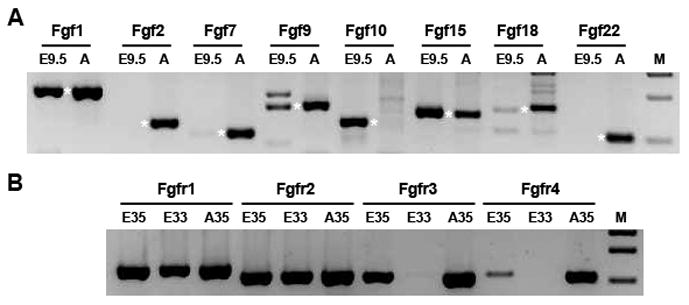
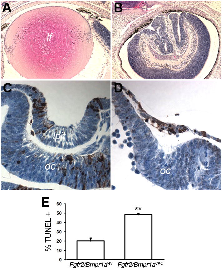
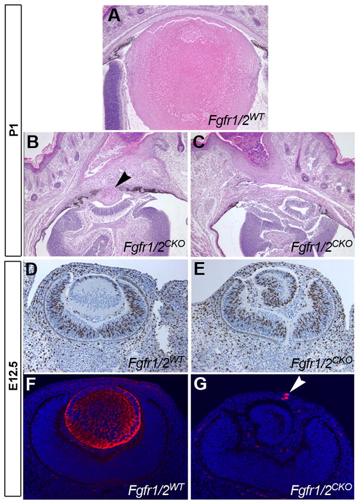
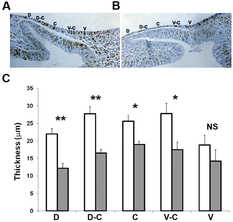
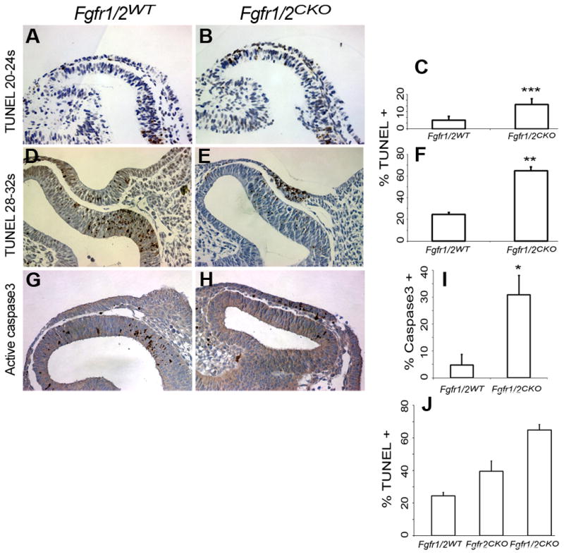
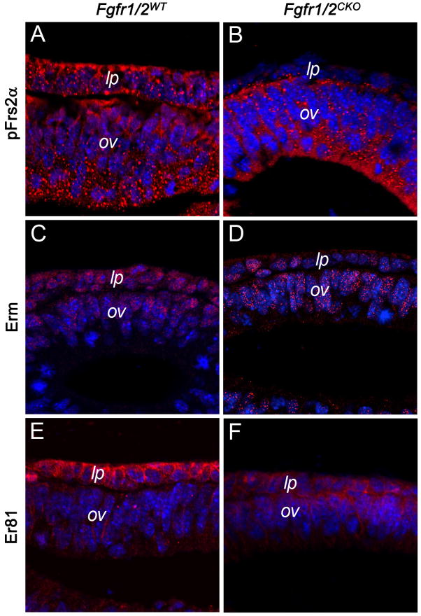
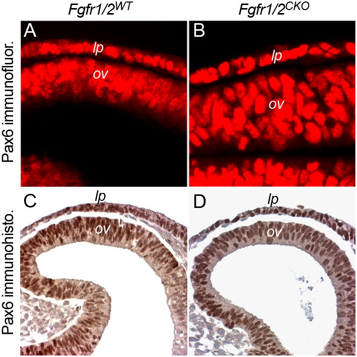
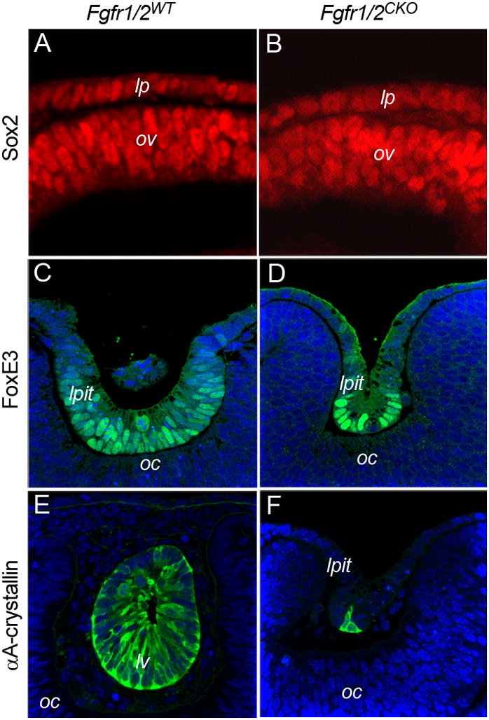
Previous studies suggested that FGF signaling is important for lens formation. However, the times at which FGFs act to promote lens formation, the FGFs that are involved, the cells that secrete them and the mechanisms by which FGF signaling may promote lens formation are not known. We found that transcripts encoding several FGF ligands and the four classical FGF receptors are detectable in the lens-forming ectoderm at the time of lens induction. Conditional deletion of Fgfr1 and Fgfr2 from this tissue resulted in the formation of small lens rudiments that soon degenerated. Lens placodes lacking Fgfr1 and 2 were thinner than in wild-type embryos. Deletion of Fgfr2 increased cell death from the initiation of placode formation and concurrent deletion of Fgfr1 enhanced this phenotype. Fgfr1/2 conditional knockout placode cells expressed lower levels of proteins known to be regulated by FGF receptor signaling, but proteins known to be important for lens formation were present at normal levels in the remaining placode cells, including the transcription factors Pax6, Sox2 and FoxE3 and the lens-preferred protein αA-crystallin. Previous studies identified a genetic interaction between BMP and FGF signaling in lens formation and conditional deletion of Bmpr1a caused increased cell death in the lens placode, resulting in the formation of smaller lenses. In the present study, conditional deletion of both Bmpr1a and Fgfr2 increased cell death beyond that seen in Fgfr2(CKO) placodes and prevented lens formation. These results suggest that the primary role of autocrine or paracrine FGF signaling is to provide essential survival signals to lens placode cells. Because apoptosis was already increased at the onset of placode formation in Fgfr1/2 conditional knockout placode cells, FGF signaling was functionally absent during the period of lens induction by the optic vesicle. Since the expression of proteins required for lens formation was not altered in the knockout placode cells, we can conclude that FGF signaling from the optic vesicle is not required for lens induction.
先前的研究表明,FGF 信号对晶状体形成很重要。然而,FGF 促进晶状体形成的作用时间、涉及的 FGF、分泌它们的细胞以及 FGF 信号促进晶状体形成的机制尚不清楚。我们发现,在晶状体诱导时,编码几种 FGF 配体和四个经典 FGF 受体的转录本可在晶状体形成外胚层中检测到。从该组织中条件性缺失 Fgfr1 和 Fgfr2 会导致小晶状体原基的形成,这些晶状体原基很快就退化了。缺乏 Fgfr1 和 2 的晶状体嵴比野生型胚胎中的更薄。从基板形成开始,Fgfr2 的缺失增加了细胞死亡,而 Fgfr1 的同时缺失增强了这种表型。Fgfr1/2 条件性敲除基板细胞表达的已知受 FGF 受体信号调节的蛋白水平较低,但在剩余的基板细胞中存在正常水平的对晶状体形成很重要的蛋白,包括转录因子 Pax6、Sox2 和 FoxE3 以及晶状体首选蛋白αA-晶体蛋白。先前的研究确定了 BMP 和 FGF 信号在晶状体形成中的遗传相互作用,并且 Bmpr1a 的条件缺失导致晶状体基板中的细胞死亡增加,从而导致较小的晶状体形成。在本研究中,Fgfr2 和 Bmpr1a 的条件缺失都增加了细胞死亡,超过了 Fgfr2(CKO)基板中看到的情况,并阻止了晶状体的形成。这些结果表明,自分泌或旁分泌 FGF 信号的主要作用是为晶状体基板细胞提供必需的存活信号。由于 Fgfr1/2 条件性敲除基板细胞中的细胞凋亡在基板形成时已经增加,因此在视泡诱导晶状体形成期间,FGF 信号在功能上缺失。由于缺失基板细胞中形成晶状体所需的蛋白表达没有改变,因此我们可以得出结论,来自视泡的 FGF 信号对于晶状体诱导不是必需的。