Moisio Kirsten, Chang Alison, Eckstein Felix, Chmiel Joan S, Wirth Wolfgang, Almagor Orit, Prasad Pottumarthi, Cahue September, Kothari Ami, Sharma Leena
Feinberg School of Medicine, Northwestern University, Chicago, IL, USA.
Arthritis Rheum. 2011 Apr;63(4):1002-9. doi: 10.1002/art.30216.
Varus-valgus alignment has been linked to subsequent progression of osteoarthritis (OA) within the mechanically stressed (medial for varus, lateral for valgus) tibiofemoral compartment. Cartilage data from the off-loaded compartment are sparse. The purpose of this study was to examine our hypotheses that neutral and valgus (versus varus) knees each have reduced odds of cartilage loss in the medial subregions and that neutral and varus (versus valgus) knees each have reduced odds of cartilage loss in the lateral subregions.
Patients with knee OA underwent knee magnetic resonance imaging at baseline and 2 years. The mean cartilage thickness was quantified within 5 tibial and 3 femoral subregions. We used logistic regression with generalized estimating equations to analyze the relationship between baseline alignment and subregional cartilage loss at 2 years, adjusting for age, sex, body mass index, and disease severity.
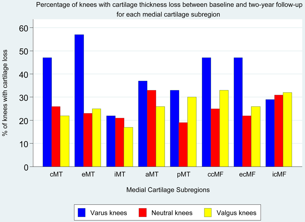
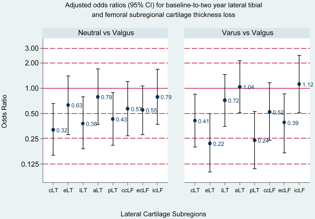
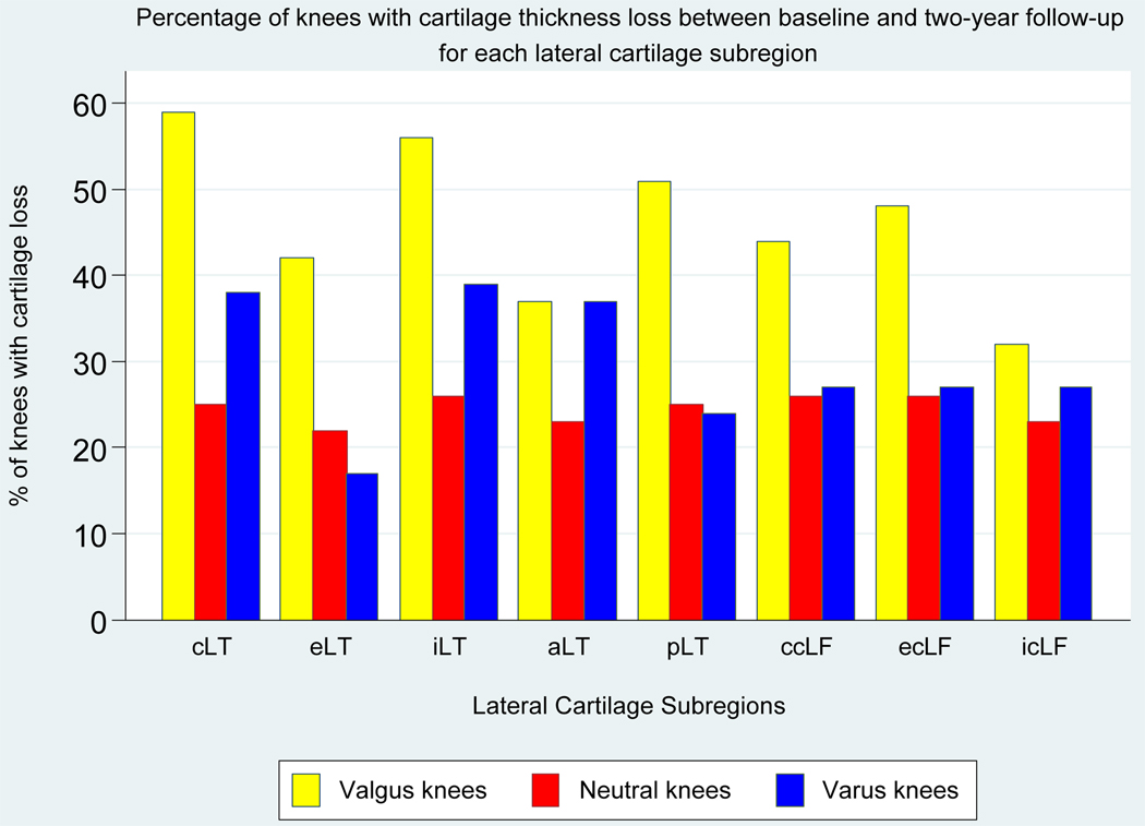
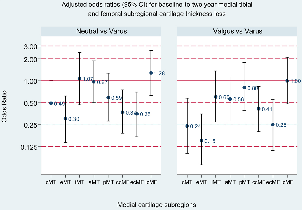
A reduced risk of cartilage loss in the medial subregions was associated with neutral (versus varus) alignment (external tibial, central femoral, external femoral) and with valgus (versus varus) alignment (central tibial, external tibial, central femoral, external femoral). A reduced risk of cartilage loss in the lateral subregions was associated with neutral (versus valgus) alignment (central tibial, internal tibial, posterior tibial) and with varus (versus valgus) alignment (central tibial, external tibial, posterior tibial, external femoral).
Neutral and valgus alignment were each associated with a reduction in the risk of subsequent cartilage loss in certain medial subregions and neutral and varus alignment with a reduction in the risk of cartilage loss in certain lateral subregions. These results support load redistribution as an in vivo mechanism of the long-term alignment effects on cartilage loss in knee OA.
膝内翻-外翻对线与机械应力作用的胫股关节间室(膝内翻时为内侧,膝外翻时为外侧)骨关节炎(OA)的后续进展相关。非负重间室的软骨数据较少。本研究的目的是检验我们的假设,即中立位和外翻(相对于内翻)膝关节在内侧亚区域软骨丢失的几率均降低,以及中立位和内翻(相对于外翻)膝关节在外侧亚区域软骨丢失的几率均降低。
膝骨关节炎患者在基线和2年时接受膝关节磁共振成像检查。在5个胫骨亚区域和3个股骨亚区域内对平均软骨厚度进行量化。我们使用广义估计方程的逻辑回归分析基线对线与2年时亚区域软骨丢失之间的关系,并对年龄、性别、体重指数和疾病严重程度进行校正。
在内侧亚区域,软骨丢失风险降低与中立位(相对于内翻)对线(胫骨外侧、股骨中央、股骨外侧)以及外翻(相对于内翻)对线(胫骨中央、胫骨外侧、股骨中央、股骨外侧)相关。在外侧亚区域,软骨丢失风险降低与中立位(相对于外翻)对线(胫骨中央、胫骨内侧、胫骨后侧)以及内翻(相对于外翻)对线(胫骨中央、胫骨外侧、胫骨后侧、股骨外侧)相关。
中立位和外翻对线均与某些内侧亚区域后续软骨丢失风险降低相关,中立位和内翻对线与某些外侧亚区域软骨丢失风险降低相关。这些结果支持负荷再分配作为膝骨关节炎中长期对线对软骨丢失影响的一种体内机制。