Department of Biological Sciences, Section of Evolutionary and Comparative Biology, University Federico II of Naples, Italy.
Eur J Histochem. 2010 Nov 5;54(4):e45. doi: 10.4081/ejh.2010.e45.
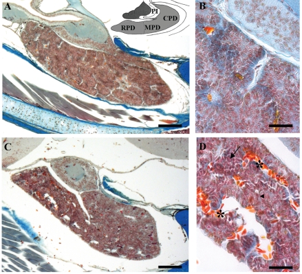
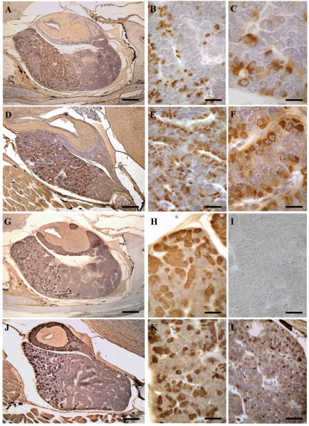
We analyzed the effect of cadmium on corticotropic (ACTH) and prolactin (PRL) cells in the pituitary gland of the Podarcis sicula lizard under chronic exposure to this metal. Adult lizards were given CdCl2 in drinking water at the dose of 10 μg/10 g body mass for 120 days. Light microscopy was performed after histological and immunohistochemical staining, and the effects were followed at regular time intervals up to 120 days post-treatment. We detected substantial variations in the general morphology of the pituitary: unlike the control lizards in which the gland appeared compact, the treated lizards showed a glandular tissue with dilated spaces that were more extensive at 90 and 120 days. PRL and ACTH cells showed an increase in occurrence and immunostaining intensity in treated lizards in comparison with the same cells of control animals. This cellular increase peaked for PRL at 30 days in the rostral, medial and also caudal pars distalis of the gland. ACTH cells appeared to increase markedly after 60 days of treatment in both the pars distalis and the pars intermedia. Again, at 60 days small, isolated ACTH cells were also found in the caudal pars distalis in which these cells were generally absent. However, at 120 days both these cellular types showed an occurrence, distribution and morphology similar to those observed in the control lizards. In lizards, protracted oral exposure to cadmium evidently involves an alteration of the normal morphology of the gland and an inhibitory effect of ACTH and PRL cells, since they increase in occurrence and immunostaining. Yet in time the inhibitory effect of cadmium on ACTH and PRL cells falls back and their occurrence appears similar to that of the control lizard.
我们分析了慢性暴露于镉对蜥蜴下丘脑中促肾上腺皮质激素(ACTH)和催乳素(PRL)细胞的影响。成年蜥蜴通过饮用水给予 CdCl2,剂量为 10μg/10g 体重,持续 120 天。在组织学和免疫组织化学染色后进行了显微镜检查,并在治疗后定期进行随访,时间长达 120 天。我们检测到垂体的一般形态发生了实质性变化:与对照组蜥蜴的致密腺组织不同,治疗组蜥蜴的腺组织出现了扩张的空间,在 90 和 120 天时更为广泛。与对照组动物的相同细胞相比,PRL 和 ACTH 细胞在治疗组蜥蜴中的出现和免疫染色强度增加。PRL 细胞的这种细胞增加在治疗后 30 天在腺的前、中、尾部表现最为明显。ACTH 细胞在治疗后 60 天在垂体前叶和垂体中间部明显增加。同样,在 60 天时,在通常没有这些细胞的尾部垂体前叶中也发现了孤立的小 ACTH 细胞。然而,在 120 天时,这两种细胞类型的出现、分布和形态都与对照组蜥蜴相似。在蜥蜴中,长期口服暴露于镉显然涉及到腺正常形态的改变,以及 ACTH 和 PRL 细胞的抑制作用,因为它们的出现和免疫染色增加。然而,随着时间的推移,镉对 ACTH 和 PRL 细胞的抑制作用减弱,其出现与对照组蜥蜴相似。