Institute of Biomedicine, University of Gothenburg, Gothenburg, Sweden.
PLoS One. 2011 Jan 10;6(1):e15846. doi: 10.1371/journal.pone.0015846.
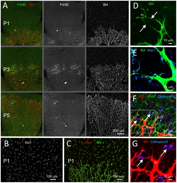
Myeloid cells have been associated with physiological and pathological angiogenesis, but their exact functions in these processes remain poorly defined. Monocyte-derived tissue macrophages of the CNS, or microglial cells, invade the mammalian retina before it becomes vascularized. Recent studies correlate the presence of microglia in the developing CNS with vascular network formation, but it is not clear whether the effect is directly caused by microglia and their contact with the endothelium.
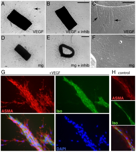
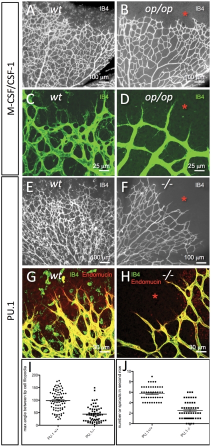
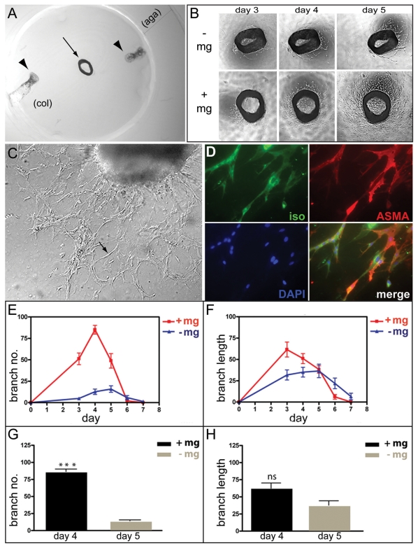
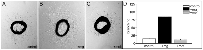
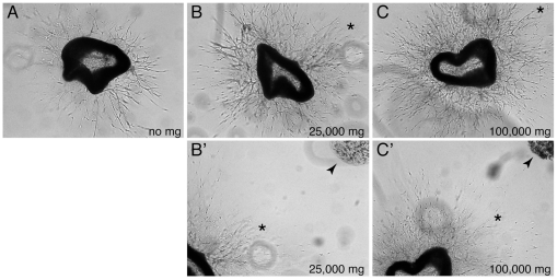
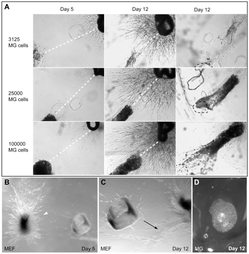
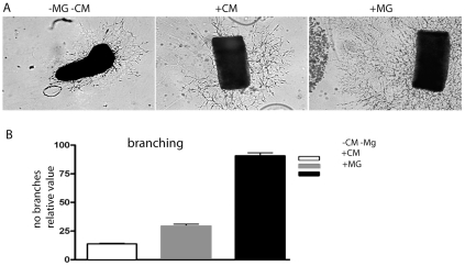
METHODOLOGY/PRINCIPAL FINDINGS: We combined in vivo studies of the developing mouse retina with in vitro studies using the aortic ring model to address the role of microglia in developmental angiogenesis. Our in vivo analyses are consistent with previous findings that microglia are present at sites of endothelial tip-cell anastomosis, and genetic ablation of microglia caused a sparser vascular network associated with reduced number of filopodia-bearing sprouts. Addition of microglia in the aortic ring model was sufficient to stimulate vessel sprouting. The effect was independent of physical contact between microglia and endothelial cells, and could be partly mimicked using microglial cell-conditioned medium. Addition of VEGF-A promoted angiogenic sprouts of different morphology in comparison with the microglial cells, and inhibition of VEGF-A did not affect the microglia-induced angiogenic response, arguing that the proangiogenic factor(s) released by microglia is distinct from VEGF-A. Finally, microglia exhibited oriented migration towards the vessels in the aortic ring cultures.
CONCLUSIONS/SIGNIFICANCE: Microglia stimulate vessel sprouting in the aortic ring cultures via a soluble microglial-derived product(s), rather than direct contact with endothelial cells. The observed migration of microglia towards the growing sprouts suggests that their position near endothelial tip-cells could result from attractive cues secreted by the vessels. Our data reveals a two-way communication between microglia and vessels that depends on soluble factors and should extend the understanding of how microglia promote vascular network formation.
髓样细胞与生理和病理血管生成有关,但它们在这些过程中的确切功能仍未得到明确界定。哺乳动物视网膜在血管化之前,单核细胞衍生的中枢神经系统组织巨噬细胞或小胶质细胞就会侵入。最近的研究将小胶质细胞在发育中的中枢神经系统中的存在与血管网络的形成相关联,但尚不清楚这种影响是否是由小胶质细胞及其与内皮细胞的接触直接引起的。
方法/主要发现:我们将发育中的小鼠视网膜的体内研究与主动脉环模型的体外研究相结合,以解决小胶质细胞在发育性血管生成中的作用。我们的体内分析与以前的发现一致,即小胶质细胞存在于内皮细胞尖端细胞吻合处,小胶质细胞的基因缺失导致血管网络稀疏,与具有更少的生芽突起的突起相关联。在主动脉环模型中添加小胶质细胞足以刺激血管发芽。该效果独立于小胶质细胞和内皮细胞之间的物理接触,并且可以使用小胶质细胞条件培养基部分模拟。与小胶质细胞相比,添加 VEGF-A 促进了不同形态的血管生成芽,并且抑制 VEGF-A 不影响小胶质细胞诱导的血管生成反应,这表明小胶质细胞释放的促血管生成因子与 VEGF-A 不同。最后,小胶质细胞在主动脉环培养物中表现出朝向血管的定向迁移。
结论/意义:小胶质细胞通过可溶性小胶质细胞衍生产物刺激主动脉环培养物中的血管发芽,而不是与内皮细胞直接接触。观察到小胶质细胞朝向生长芽的迁移表明,它们在内皮细胞尖端细胞附近的位置可能是由于血管分泌的吸引信号所致。我们的数据揭示了小胶质细胞和血管之间的双向通讯,这取决于可溶性因子,应该扩展对小胶质细胞如何促进血管网络形成的理解。