Department of Nuclear Medicine & PET Research, VU University Medical Center, PO Box 7057, 1007 MB Amsterdam, The Netherlands.
Eur J Nucl Med Mol Imaging. 2011 May;38(5):930-9. doi: 10.1007/s00259-011-1730-3. Epub 2011 Jan 27.
Parametric imaging of absolute myocardial blood flow (MBF) using [(15)O]H(2)O enables determination of MBF with high spatial resolution. The aim of this study was to develop a method for generating reproducible, high-quality and quantitative parametric MBF images with minimal user intervention.
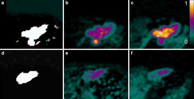
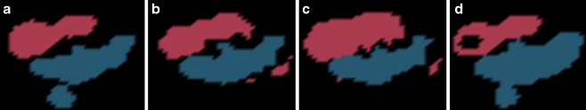
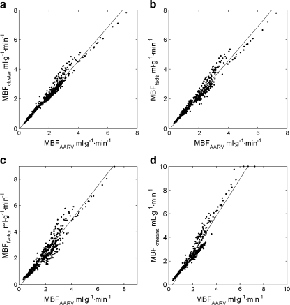
Nineteen patients referred for evaluation of MBF underwent rest and adenosine stress [(15)O]H(2)O positron emission tomography (PET) scans. Ascending aorta and right ventricular (RV) cavity volumes of interest (VOIs) were used as input functions. Implementation of a basis function method (BFM) of the single-tissue model with an additional correction for RV spillover was used to generate parametric images. The average segmental MBF derived from parametric images was compared with MBF obtained using nonlinear least-squares regression (NLR) of VOI data. Four segmentation algorithms were evaluated for automatic extraction of input functions. Segmental MBF obtained using these input functions was compared with MBF obtained using manually defined input functions.
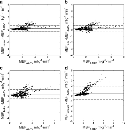
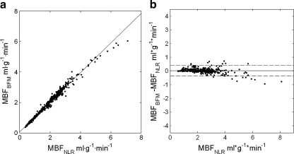
The average parametric MBF showed a high agreement with NLR-derived MBF [intraclass correlation coefficient (ICC) = 0.984]. For each segmentation algorithm there was at least one implementation that yielded high agreement (ICC > 0.9) with manually obtained input functions, although MBF calculated using each algorithm was at least 10% higher. Cluster analysis with six clusters yielded the highest agreement (ICC = 0.977), together with good segmentation reproducibility (coefficient of variation of MBF <5%).
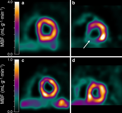
Parametric MBF images of diagnostic quality can be generated automatically using cluster analysis and a implementation of a BFM of the single-tissue model with additional RV spillover correction.
使用 [(15)O]H(2)O 对绝对心肌血流 (MBF) 进行参数成像,可实现 MBF 的高空间分辨率测定。本研究旨在开发一种方法,以最少的用户干预生成可重复、高质量和定量的参数 MBF 图像。
19 例因 MBF 评估而转诊的患者接受了静息和腺苷负荷 [(15)O]H(2)O 正电子发射断层扫描 (PET) 扫描。升主动脉和右心室 (RV) 腔容积感兴趣区 (VOI) 被用作输入函数。采用单组织模型的基函数方法 (BFM) 并对 RV 溢出进行额外校正,以生成参数图像。从参数图像中得出的平均节段性 MBF 与使用 VOI 数据的非线性最小二乘回归 (NLR) 获得的 MBF 进行比较。评估了四种分割算法,以自动提取输入函数。将使用这些输入函数获得的节段性 MBF 与使用手动定义的输入函数获得的 MBF 进行比较。
平均参数性 MBF 与 NLR 衍生的 MBF 高度一致 [组内相关系数 (ICC) = 0.984]。对于每种分割算法,至少有一种实现与手动获得的输入函数具有高度一致性 (ICC > 0.9),尽管使用每种算法计算的 MBF 至少高 10%。使用聚类分析的 6 个聚类产生了最高的一致性 (ICC = 0.977),同时具有良好的分割可重复性 (MBF 变异系数 <5%)。
使用聚类分析和单组织模型的 BFM 与 RV 溢出校正的实现,可以自动生成诊断质量的参数性 MBF 图像。