Institute of Medical Science, Faculty of Medicine, University of Toronto, Toronto, ON, Canada.
Respir Res. 2011 Feb 3;12(1):19. doi: 10.1186/1465-9921-12-19.
Arginase overexpression contributes to airways hyperresponsiveness (AHR) in asthma. Arginase expression is further augmented in cigarette smoking asthmatics, suggesting that it may be upregulated by environmental pollution. Thus, we hypothesize that arginase contributes to the exacerbation of respiratory symptoms following exposure to air pollution, and that pharmacologic inhibition of arginase would abrogate the pollution-induced AHR.
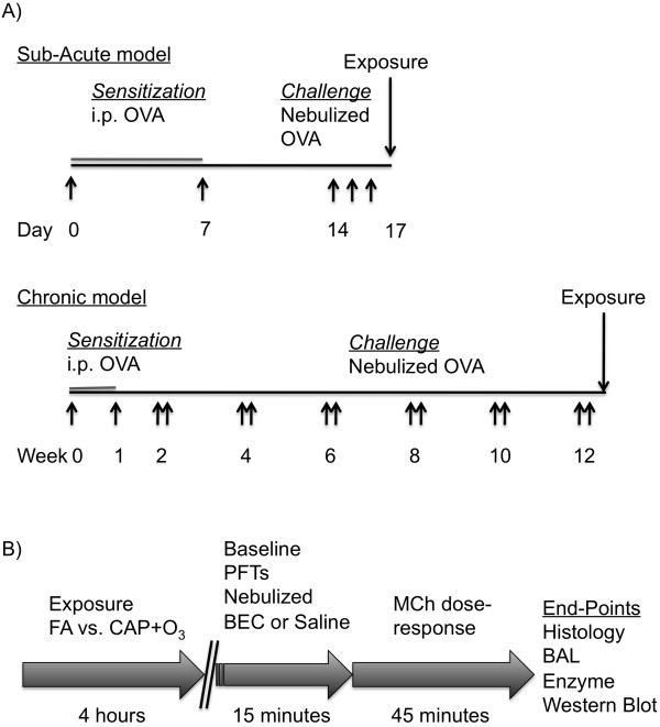
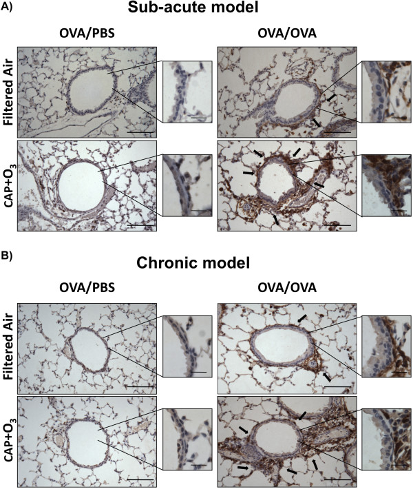
To investigate the role of arginase in the air pollution-induced exacerbation of airways responsiveness, we employed two murine models of allergic airways inflammation. Mice were sensitized to ovalbumin (OVA) and challenged with nebulized PBS (OVA/PBS) or OVA (OVA/OVA) for three consecutive days (sub-acute model) or 12 weeks (chronic model), which exhibit inflammatory cell influx and remodeling/AHR, respectively. Twenty-four hours after the final challenge, mice were exposed to concentrated ambient fine particles plus ozone (CAP+O₃), or HEPA-filtered air (FA), for 4 hours. After the CAP+O₃ exposures, mice underwent tracheal cannulation and were treated with an aerosolized arginase inhibitor (S-boronoethyl-L-cysteine; BEC) or vehicle, immediately before determination of respiratory function and methacholine-responsiveness using the flexiVent®. Lungs were then collected for comparison of arginase activity, protein expression, and immunohistochemical localization.
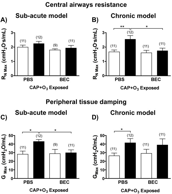
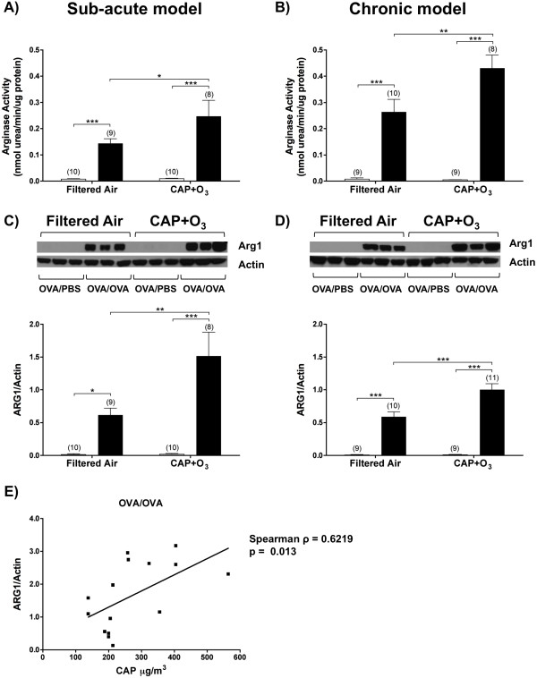
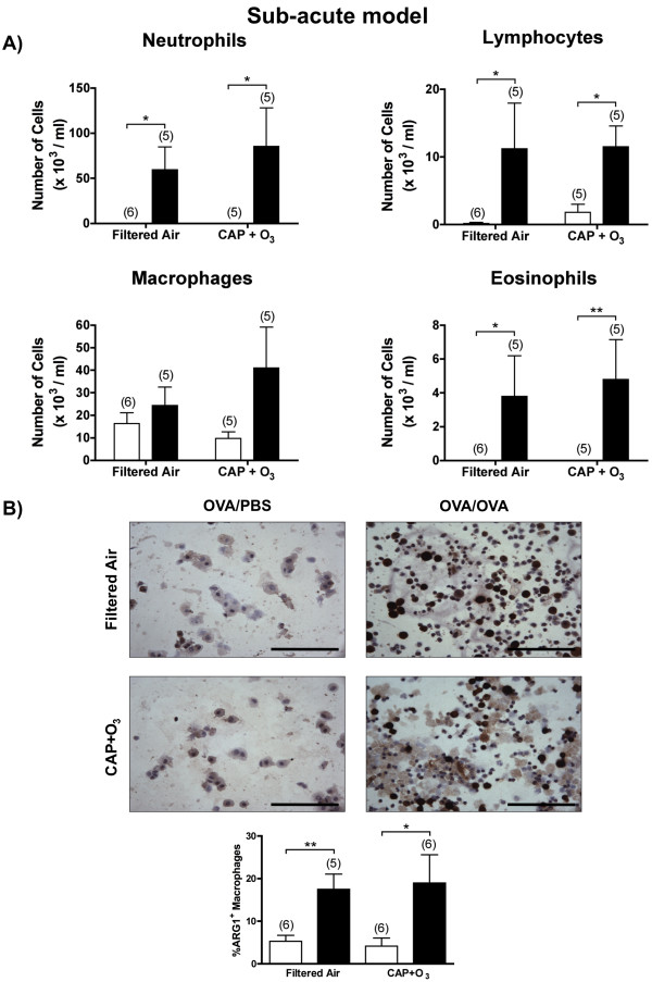
Compared to FA, arginase activity was significantly augmented in the lungs of CAP+O₃-exposed OVA/OVA mice in both the sub-acute and chronic models. Western blotting and immunohistochemical staining revealed that the increased activity was due to arginase 1 expression in the area surrounding the airways in both models. Arginase inhibition significantly reduced the CAP+O₃-induced increase in AHR in both models.
This study demonstrates that arginase is upregulated following environmental exposures in murine models of asthma, and contributes to the pollution-induced exacerbation of airways responsiveness. Thus arginase may be a therapeutic target to protect susceptible populations against the adverse health effects of air pollution, such as fine particles and ozone, which are two of the major contributors to smog.
精氨酸酶过表达导致哮喘患者气道高反应性(AHR)。在吸烟的哮喘患者中,精氨酸酶的表达进一步增加,这表明它可能受到环境污染的上调。因此,我们假设精氨酸酶导致暴露于空气污染后呼吸症状的恶化,并且精氨酸酶的药物抑制会消除污染引起的 AHR。
为了研究精氨酸酶在空气污染引起的气道反应性恶化中的作用,我们采用了两种变应性气道炎症的小鼠模型。小鼠用卵清蛋白(OVA)致敏,并连续 3 天(亚急性模型)或 12 周(慢性模型)用雾化 PBS(OVA/PBS)或 OVA(OVA/OVA)进行雾化挑战,分别表现为炎症细胞浸润和重塑/AHR。最后一次挑战后 24 小时,小鼠暴露于浓缩环境细颗粒物加臭氧(CAP+O₃)或高效空气过滤器(FA)中 4 小时。暴露于 CAP+O₃ 后,小鼠进行气管插管,并在 FlexiVent®上测定呼吸功能和乙酰甲胆碱反应性之前,用雾化精氨酸酶抑制剂(S-硼代乙基-L-半胱氨酸;BEC)或载体处理。然后收集肺比较精氨酸酶活性、蛋白表达和免疫组织化学定位。
与 FA 相比,在亚急性和慢性模型中,CAP+O₃ 暴露的 OVA/OVA 小鼠的肺中精氨酸酶活性显著增加。Western 印迹和免疫组织化学染色显示,在两种模型中,活性的增加是由于气道周围区域的精氨酸酶 1 表达增加所致。精氨酸酶抑制显著降低了两种模型中 CAP+O₃ 引起的 AHR 增加。
本研究表明,在哮喘小鼠模型中,环境暴露后精氨酸酶上调,并导致气道反应性对污染的加剧。因此,精氨酸酶可能是一种治疗靶点,以保护易感人群免受空气污染(如细颗粒物和臭氧)等不良健康影响,这两种污染物是烟雾的主要贡献者之一。