State Key Laboratory for Infectious Disease Prevention and Control, National Institute for Viral Disease Control and Prevention, Chinese Center for Disease Control and Prevention, Beijing, People's Republic of China.
PLoS One. 2011 Jan 27;6(1):e14602. doi: 10.1371/journal.pone.0014602.
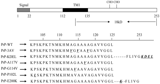
Genetic prion diseases are linked to point and inserted mutations in the prion protein (PrP) gene that are presumed to favor conversion of the cellular isoform of PrP (PrP(C)) to the pathogenic one (PrP(Sc)). The pathogenic mechanisms and the subcellular sites of the conversion are not completely understood. Here we introduce several PRNP gene mutations (such as, PrP-KDEL, PrP-3AV, PrP-A117V, PrP-G114V, PrP-P102L and PrP-E200K) into the cultured cells in order to explore the pathogenic mechanism of familial prion disease.
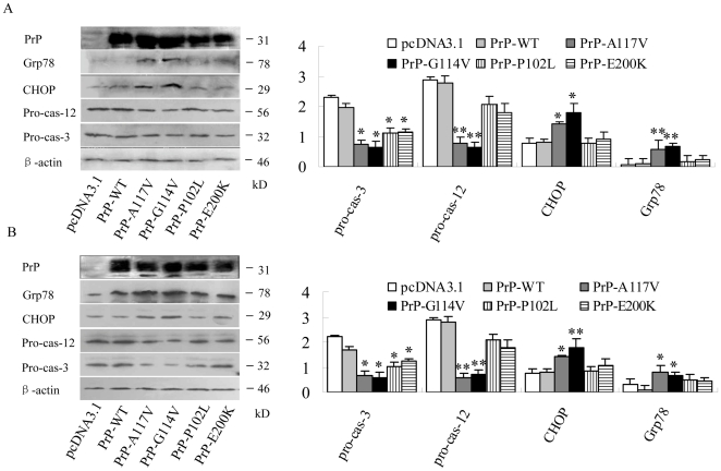
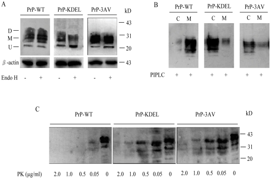
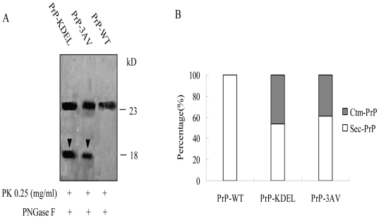
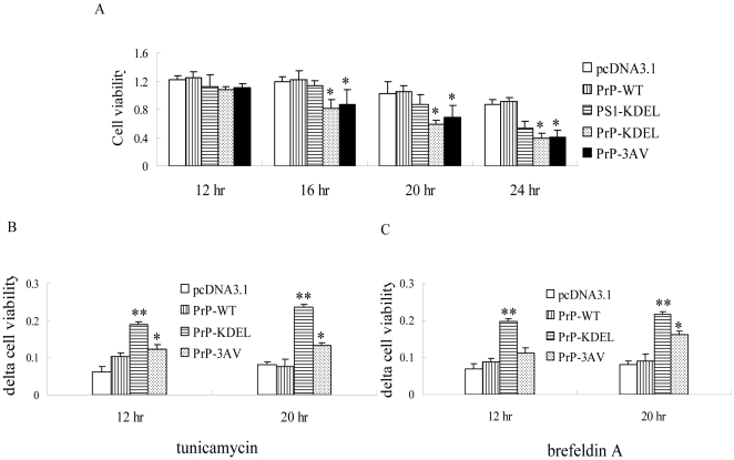
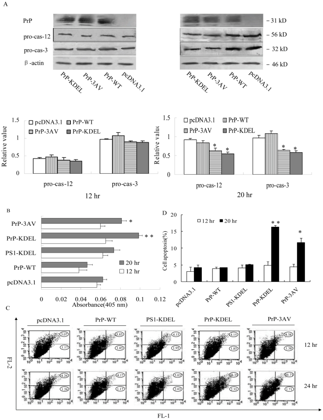
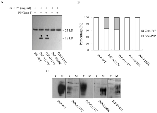
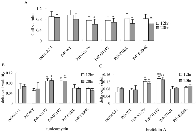
METHODOLOGY/PRINCIPAL FINDINGS: To address the roles of aberrant retention of PrP in endoplasmic reticulum (ER), the recombinant plasmids expressing full-length human PrP tailed with an ER signal peptide at the COOH-terminal (PrP-KDEL) and PrP with three amino acids exchange in transmembrane region (PrP-3AV) were constructed. In the preparations of transient transfections, 18-kD COOH-terminal proteolytic resistant fragments (Ctm-PrP) were detected in the cells expressing PrP-KDEL and PrP-3AV. Analyses of the cell viabilities in the presences of tunicamycin and brefeldin A revealed that expressions of PrP-KDEL and PrP-3AV sensitized the transfected cells to ER stress stimuli. Western blots and RT-PCR identified the clear alternations of ER stress associated events in the cells expressing PrP-KDEL and PrP-3AV that induced ER mediated apoptosis by CHOP and caspase-12 apoptosis pathway. Moreover, several familial CJD related PrP mutants were transiently introduced into the cultured cells. Only the mutants within the transmembrane region (G114V and A117V) induced the formation of Ctm-PrP and caused the ER stress, while the mutants outside the transmembrane region (P102L and E200K) failed.
CONCLUSIONS/SIGNIFICANCE: The data indicate that the retention of PrP in ER through formation of Ctm-PrP results in ER stress and cell apoptosis. The cytopathic activities caused by different familial CJD associated PrP mutants may vary, among them the mutants within the transmembrane region undergo an ER-stress mediated cell apoptosis.
遗传朊病毒疾病与朊病毒蛋白(PrP)基因中的点突变和插入突变有关,这些突变被认为有利于细胞型 PrP(PrP(C))向致病性 PrP(PrP(Sc))的转化。转化的致病机制和亚细胞部位尚不完全清楚。在这里,我们将几种 PRNP 基因突变(如 PrP-KDEL、PrP-3AV、PrP-A117V、PrP-G114V、PrP-P102L 和 PrP-E200K)引入培养细胞中,以探索家族性朊病毒病的致病机制。
方法/主要发现:为了研究 PrP 在内质网(ER)中的异常滞留在致病机制中的作用,构建了在 COOH 末端带有 ER 信号肽的全长人 PrP(PrP-KDEL)和在跨膜区有三个氨基酸置换的 PrP(PrP-3AV)的重组质粒。在瞬时转染的制剂中,在表达 PrP-KDEL 和 PrP-3AV 的细胞中检测到 18-kD COOH 末端蛋白酶抗性片段(Ctm-PrP)。在存在衣霉素和布雷菲德菌素 A 的情况下分析细胞活力的结果表明,PrP-KDEL 和 PrP-3AV 的表达使转染细胞对 ER 应激刺激敏感。Western blot 和 RT-PCR 鉴定了表达 PrP-KDEL 和 PrP-3AV 的细胞中与 ER 应激相关的事件的明显改变,这些改变通过 CHOP 和 caspase-12 凋亡途径诱导 ER 介导的细胞凋亡。此外,还将几种家族性 CJD 相关的 PrP 突变体瞬时引入培养细胞中。只有跨膜区的突变体(G114V 和 A117V)诱导 Ctm-PrP 的形成并引起 ER 应激,而跨膜区以外的突变体(P102L 和 E200K)则没有。
结论/意义:数据表明,通过形成 Ctm-PrP 将 PrP 滞留在 ER 中会导致 ER 应激和细胞凋亡。不同家族性 CJD 相关 PrP 突变体引起的细胞病变活性可能不同,其中跨膜区的突变体经历 ER 应激介导的细胞凋亡。