• 文献检索
  • 文档翻译
  • 深度研究
  • 学术资讯
  • Suppr Zotero 插件Zotero 插件
  • 邀请有礼
  • 套餐&价格
  • 历史记录
应用&插件
Suppr Zotero 插件Zotero 插件浏览器插件Mac 客户端Windows 客户端微信小程序
定价
高级版会员购买积分包购买API积分包
服务
文献检索文档翻译深度研究API 文档MCP 服务
关于我们
关于 Suppr公司介绍联系我们用户协议隐私条款
关注我们

Suppr 超能文献

核心技术专利:CN118964589B侵权必究
粤ICP备2023148730 号-1Suppr @ 2026

文献检索

告别复杂PubMed语法,用中文像聊天一样搜索,搜遍4000万医学文献。AI智能推荐,让科研检索更轻松。

立即免费搜索

文件翻译

保留排版,准确专业,支持PDF/Word/PPT等文件格式,支持 12+语言互译。

免费翻译文档

深度研究

AI帮你快速写综述,25分钟生成高质量综述,智能提取关键信息,辅助科研写作。

立即免费体验

颅内镍钛诺支架的高分辨率 3D X 射线成像。

High-resolution 3D X-ray imaging of intracranial nitinol stents.

机构信息

Faculty Electrical Engineering, Signal Processing Systems group (SPS), Eindhoven University of Technology (TU/e), Laplace Building 028, Postbox 513, 5600MB, Eindhoven, The Netherlands.

出版信息

Neuroradiology. 2012 Feb;54(2):155-62. doi: 10.1007/s00234-011-0839-1. Epub 2011 Feb 18.

DOI:10.1007/s00234-011-0839-1
PMID:21331601
原文链接:https://pmc.ncbi.nlm.nih.gov/articles/PMC3261414/
Abstract

INTRODUCTION

To assess an optimized 3D imaging protocol for intracranial nitinol stents in 3D C-arm flat detector imaging. For this purpose, an image quality simulation and an in vitro study was carried out.

METHODS

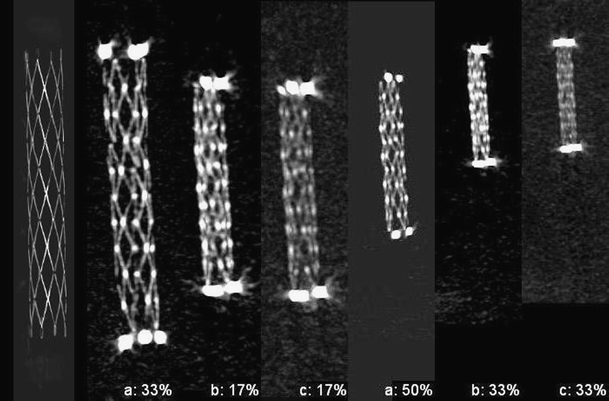
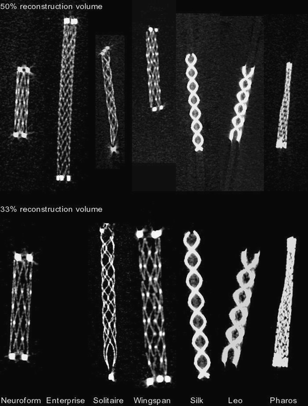
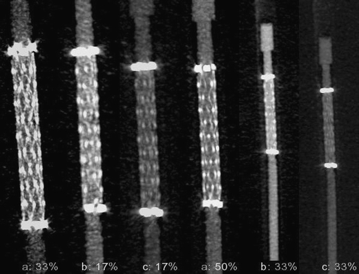
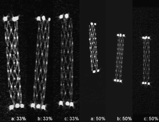
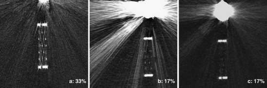
Nitinol stents of various brands were placed inside an anthropomorphic head phantom, using iodine contrast. Experiments with objects were preceded by image quality and dose simulations. We varied X-ray imaging parameters in a commercially interventional X-ray system to set 3D image quality in the contrast-noise-sharpness space. Beam quality was varied to evaluate contrast of the stents while keeping absorbed dose below recommended values. Two detector formats were used, paired with an appropriate pixel size and X-ray focus size. Zoomed reconstructions were carried out and snapshot images acquired. High contrast spatial resolution was assessed with a CT phantom.

RESULTS

We found an optimal protocol for imaging intracranial nitinol stents. Contrast resolution was optimized for nickel-titanium-containing stents. A high spatial resolution larger than 2.1 lp/mm allows struts to be visualized. We obtained images of stents of various brands and a representative set of images is shown. Independent of the make, struts can be imaged with virtually continuous strokes. Measured absorbed doses are shown to be lower than 50 mGy Computed Tomography Dose Index (CTDI).

CONCLUSION

By balancing the modulation transfer of the imaging components and tuning the high-contrast imaging capabilities, we have shown that thin nitinol stent wires can be reconstructed with high contrast-to-noise ratio and good detail, while keeping radiation doses within recommended values. Experimental results compare well with imaging simulations.

摘要

简介

评估 3D C 臂平板探测器成像中颅内镍钛合金支架的优化 3D 成像方案。为此,进行了图像质量模拟和体外研究。

方法

将各种品牌的镍钛合金支架用碘对比剂放置在人体头部模型内。在进行物体实验之前,进行了图像质量和剂量模拟。我们在商业介入 X 射线系统中改变 X 射线成像参数,以在对比度-噪声-锐度空间中设置 3D 图像质量。改变射线质量以评估支架的对比度,同时将吸收剂量保持在推荐值以下。使用两种探测器格式,与适当的像素大小和 X 射线焦点大小相匹配。进行了缩放重建并获取了快照图像。使用 CT 体模评估高对比度空间分辨率。

结果

我们找到了一种优化的颅内镍钛合金支架成像方案。对于含镍钛的支架,优化了对比度分辨率。高空间分辨率大于 2.1lp/mm 可使支架的支柱可视化。我们获得了各种品牌的支架图像,并展示了一组有代表性的图像。无论品牌如何,都可以使用几乎连续的笔划对支柱进行成像。测量的吸收剂量显示低于 50mGy 计算机断层扫描剂量指数(CTDI)。

结论

通过平衡成像组件的调制传递和调整高对比度成像能力,我们表明可以用高对比度噪声比和良好的细节重建薄的镍钛合金支架线,同时将辐射剂量保持在推荐值内。实验结果与成像模拟吻合良好。

https://cdn.ncbi.nlm.nih.gov/pmc/blobs/462c/3261414/22089e6970a6/234_2011_839_Fig5_HTML.jpg
https://cdn.ncbi.nlm.nih.gov/pmc/blobs/462c/3261414/60ef264f9782/234_2011_839_Fig1_HTML.jpg
https://cdn.ncbi.nlm.nih.gov/pmc/blobs/462c/3261414/d7d2a4033ab0/234_2011_839_Fig2_HTML.jpg
https://cdn.ncbi.nlm.nih.gov/pmc/blobs/462c/3261414/856e9e952d9b/234_2011_839_Fig3_HTML.jpg
https://cdn.ncbi.nlm.nih.gov/pmc/blobs/462c/3261414/e508206cbfcf/234_2011_839_Fig4_HTML.jpg
https://cdn.ncbi.nlm.nih.gov/pmc/blobs/462c/3261414/22089e6970a6/234_2011_839_Fig5_HTML.jpg

相似文献

1
High-resolution 3D X-ray imaging of intracranial nitinol stents.颅内镍钛诺支架的高分辨率 3D X 射线成像。
Neuroradiology. 2012 Feb;54(2):155-62. doi: 10.1007/s00234-011-0839-1. Epub 2011 Feb 18.
2
Volume-of-interest (VOI) imaging in C-arm flat-detector CT for high image quality at reduced dose.C 臂平板探测器 CT 中的感兴趣区(VOI)成像,可在降低剂量的情况下获得高质量图像。
Med Phys. 2010 Jun;37(6):2719-30. doi: 10.1118/1.3427641.
3
Mobile C-arm cone-beam CT for guidance of spine surgery: image quality, radiation dose, and integration with interventional guidance.移动式 C 臂锥形束 CT 引导脊柱手术:图像质量、辐射剂量和与介入引导的整合。
Med Phys. 2011 Aug;38(8):4563-74. doi: 10.1118/1.3597566.
4
Technical assessment of a cone-beam CT scanner for otolaryngology imaging: image quality, dose, and technique protocols.用于耳鼻喉科成像的锥形束CT扫描仪的技术评估:图像质量、剂量和技术方案。
Med Phys. 2012 Aug;39(8):4932-42. doi: 10.1118/1.4736805.
5
Dual-energy CT angiography in the evaluation of intracranial aneurysms: image quality, radiation dose, and comparison with 3D rotational digital subtraction angiography.双能量 CT 血管造影在颅内动脉瘤评估中的应用:图像质量、辐射剂量,以及与三维旋转数字减影血管造影的比较。
AJR Am J Roentgenol. 2010 Jan;194(1):23-30. doi: 10.2214/AJR.08.2290.
6
Validation of synthesized normal-resolution image data generated from high-resolution acquisitions on a commercial CT scanner.在商用CT扫描仪上对由高分辨率采集生成的合成正常分辨率图像数据进行验证。
Med Phys. 2020 Oct;47(10):4775-4785. doi: 10.1002/mp.14395. Epub 2020 Aug 5.
7
Collimation and Image Quality of C-Arm Computed Tomography: Potential of Radiation Dose Reduction While Maintaining Equal Image Quality.C型臂计算机断层扫描的准直与图像质量:在保持同等图像质量的同时降低辐射剂量的潜力
Invest Radiol. 2015 Aug;50(8):514-21. doi: 10.1097/RLI.0000000000000158.
8
A mobile isocentric C-arm for intraoperative cone-beam CT: Technical assessment of dose and 3D imaging performance.术中锥形束 CT 的移动等中心 C 臂:剂量和 3D 成像性能的技术评估。
Med Phys. 2020 Mar;47(3):958-974. doi: 10.1002/mp.13983. Epub 2020 Jan 6.
9
Spatial resolution improvement and dose reduction potential for inner ear CT imaging using a z-axis deconvolution technique.利用 z 轴反卷积技术提高内耳 CT 成像的空间分辨率和降低剂量的潜力。
Med Phys. 2013 Jun;40(6):061904. doi: 10.1118/1.4802730.
10
Spatial resolution limits of multislice computed tomography (MS-CT), C-arm-CT, and flat panel-CT (FP-CT) compared to MicroCT for visualization of a small metallic stent.多层计算机断层扫描(MS-CT)、C 臂 CT 和平板 CT(FP-CT)与 MicroCT 用于显示小金属支架的空间分辨率比较。
Acad Radiol. 2011 Jul;18(7):866-75. doi: 10.1016/j.acra.2011.02.012.

引用本文的文献

1
Image Quality and Radiation Dose of Conventional and Wide-Field High-Resolution Cone-Beam Computed Tomography for Cerebral Angiography: A Phantom Study.常规和宽视野高分辨率锥形束计算机断层扫描在脑血管造影中的图像质量和辐射剂量:一项体模研究。
Tomography. 2023 Sep 1;9(5):1683-1693. doi: 10.3390/tomography9050134.
2
Investigation of Stent Prototypes for the Eustachian Tube in Human Donor Bodies.人体供体中咽鼓管支架原型的研究
Bioengineering (Basel). 2023 Jun 20;10(6):743. doi: 10.3390/bioengineering10060743.
3
Radiation dose and image quality comparison during spine surgery with two different, intraoperative 3D imaging navigation systems.

本文引用的文献

1
An in vitro study of silk stent morphology.丝支架形态的体外研究。
Neuroradiology. 2011 Sep;53(9):659-67. doi: 10.1007/s00234-010-0784-4. Epub 2010 Oct 28.
2
Contrast-enhanced angiographic cone-beam CT of cerebrovascular stents: experimental optimization and clinical application.脑血管支架的对比增强血管造影锥形束 CT:实验优化和临床应用。
AJNR Am J Neuroradiol. 2011 Jan;32(1):137-44. doi: 10.3174/ajnr.A2239. Epub 2010 Oct 21.
3
Intracranial stenting in atherosclerotic disease-recent results and challenges to face.颅内支架置入术治疗动脉粥样硬化性疾病:近期结果与面临的挑战。
使用两种不同的术中三维成像导航系统进行脊柱手术时的辐射剂量与图像质量比较。
J Appl Clin Med Phys. 2019 Feb;20(2):136-145. doi: 10.1002/acm2.12534. Epub 2019 Jan 24.
4
Usefulness of Cone Beam Intra-Arterial CTA for Evaluation of Flow Diverters: A Practical Approach for Daily Use.锥形束动脉内CT血管造影术在评估血流导向装置中的应用:日常使用的实用方法
Interv Neurol. 2018 Oct;7(6):457-463. doi: 10.1159/000490577. Epub 2018 Jul 18.
5
Role of C-arm VasoCT in the use of endovascular WEB flow disruption in intracranial aneurysm treatment.C形臂血管CT在颅内动脉瘤治疗中血管内WEB血流阻断应用中的作用。
AJNR Am J Neuroradiol. 2014 Jul;35(7):1353-7. doi: 10.3174/ajnr.A3860. Epub 2014 Feb 13.
6
Comparison of high-resolution X-ray and micro-CT for experimental evaluation of intracranial stent prototypes: quality evaluation beyond CE mark.高分辨率X射线与微型计算机断层扫描用于颅内支架原型实验评估的比较:超越CE标志的质量评估
Neuroradiology. 2014 Apr;56(4):315-23. doi: 10.1007/s00234-014-1324-4. Epub 2014 Jan 31.
7
Quantitative analysis of high-resolution, contrast-enhanced, cone-beam CT for the detection of intracranial in-stent hyperplasia.高分辨率、对比增强锥形束CT用于检测颅内支架内增生的定量分析。
J Neurointerv Surg. 2015 Feb;7(2):118-25. doi: 10.1136/neurintsurg-2013-010950. Epub 2014 Jan 30.
8
Reduction of coil mass artifacts in high-resolution flat detector conebeam CT of cerebral stent-assisted coiling.降低高分辨率平板探测器锥形束 CT 下脑动脉支架辅助弹簧圈栓塞术中的线圈伪影。
AJNR Am J Neuroradiol. 2013 Nov-Dec;34(11):2163-70. doi: 10.3174/ajnr.A3561. Epub 2013 May 30.
9
Angiographic CT: in vitro comparison of different carotid artery stents-does stent orientation matter?血管造影 CT:不同颈动脉支架的体外比较——支架方向是否重要?
Neuroradiology. 2013 Jun;55(6):675-82. doi: 10.1007/s00234-013-1152-y. Epub 2013 Feb 15.
10
Intravenous flat-detector CT angiography in acute ischemic stroke management.静脉内平板探测器 CT 血管造影在急性缺血性脑卒中治疗中的应用。
Neuroradiology. 2012 Apr;54(4):383-91. doi: 10.1007/s00234-011-0893-8. Epub 2011 May 31.
Neuroradiology. 2010 Jul;52(7):633-44. doi: 10.1007/s00234-010-0678-5. Epub 2010 Mar 30.
4
C-arm flat detector computed tomography: the technique and its applications in interventional neuro-radiology.C 臂平板探测器 CT:技术及其在介入神经放射学中的应用。
Neuroradiology. 2010 Apr;52(4):319-27. doi: 10.1007/s00234-009-0609-5. Epub 2009 Oct 27.
5
Shape memory alloys: metallurgy, biocompatibility, and biomechanics for neurosurgical applications.形状记忆合金:用于神经外科应用的冶金学、生物相容性和生物力学
Neurosurgery. 2009 May;64(5 Suppl 2):199-214; discussion 214-5. doi: 10.1227/01.NEU.0000330392.09889.99.
6
Advances in interventional neuroradiology.介入神经放射学的进展
Stroke. 2009 May;40(5):e305-12. doi: 10.1161/STROKEAHA.109.548438.
7
Endovascular occlusion of intracranial wide-necked aneurysms with stenting (Neuroform) and coiling: mid-term and long-term results.采用支架置入术(Neuroform)联合弹簧圈栓塞术对颅内宽颈动脉瘤进行血管内闭塞:中期和长期结果
Neuroradiology. 2009 Jun;51(6):401-9. doi: 10.1007/s00234-009-0502-2. Epub 2009 Feb 25.
8
Study of conformability of the new leo plus stent to a curved vascular model using flat-panel detector computed tomography (DynaCT).使用平板探测器计算机断层扫描(DynaCT)研究新型leo plus支架与弯曲血管模型的贴合性。
Neurosurgery. 2009 Mar;64(3 Suppl):ons130-4; discussion ons134. doi: 10.1227/01.NEU.0000336304.26756.21.
9
Brain imaging with a flat detector C-arm : Technique and clinical interest of XperCT.使用平板探测器C形臂进行脑部成像:XperCT的技术与临床意义
Neuroradiology. 2008 Oct;50(10):863-8. doi: 10.1007/s00234-008-0419-1. Epub 2008 Jun 17.
10
C-arm cone-beam CT: general principles and technical considerations for use in interventional radiology.C形臂锥形束CT:介入放射学应用的一般原理和技术考量
J Vasc Interv Radiol. 2008 Jun;19(6):814-20. doi: 10.1016/j.jvir.2008.02.002. Epub 2008 Apr 23.