Department of Radiology, Johns Hopkins University School of Medicine, Baltimore, MD, USA.
Semin Radiat Oncol. 2011 Apr;21(2):88-100. doi: 10.1016/j.semradonc.2010.11.004.
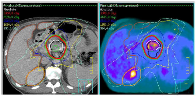
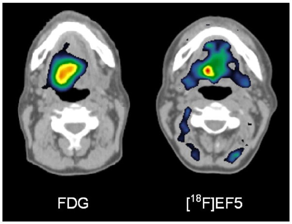
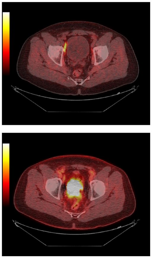
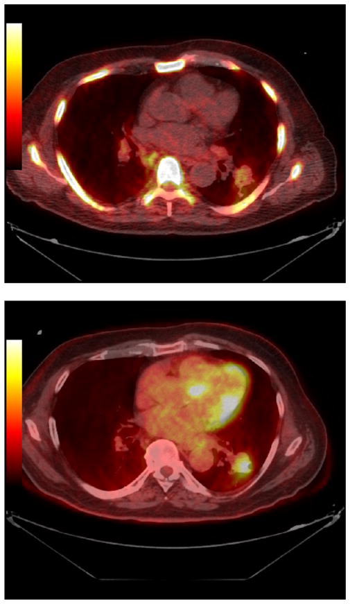
External beam radiation therapy procedures have, until recently, been planned almost exclusively using anatomic imaging methods. Molecular imaging using hybrid positron emission tomography (PET)/computed tomography scanning or single-photon emission computed tomography (SPECT) imaging has provided new insights into the precise location of tumors (staging) and the extent and character of the biologically active tumor volume (BTV) and has provided differential response information during and after therapy. In addition to the commonly used radiotracer (18)F-fluoro- 2-deoxyD-glucose (FDG), additional radiopharmaceuticals are being explored to image major physiological processes as well as tumor biological properties, such as hypoxia, proliferation, amino acid accumulation, apoptosis, and receptor expression, providing the potential to target or boost the radiation dose to a biologically relevant region within a tumor, such as the most hypoxic or most proliferative area. Imaging using SPECT agents has furthered the possibility of limiting dose to functional normal tissues. PET can also portray the distribution of particle therapy by displaying activated species in situ. With both PET and SPECT imaging, fundamental physical issues of limited spatial resolution relative to the biological process, partial volume effects for quantification of small volumes, image misregistration, motion, and edge delineation must be carefully considered and can differ by agent or the method applied. Molecular imaging-guided radiation therapy (MIGRT) is a rapidly evolving and promising area of investigation and clinical translation. As MIGRT evolves, evidence must continue to be gathered to support improved clinical outcomes using MIGRT versus purely anatomic approaches.
直到最近,外部束放射治疗程序几乎完全是使用解剖成像方法来规划的。使用正电子发射断层扫描(PET)/计算机断层扫描或单光子发射计算机断层扫描(SPECT)成像的分子成像为肿瘤的精确位置(分期)以及肿瘤生物学活性体积(BTV)的程度和特征提供了新的见解,并在治疗期间和之后提供了差异反应信息。除了常用的放射性示踪剂(18)F-氟-2-脱氧-D-葡萄糖(FDG)外,还在探索其他放射性药物来对主要生理过程以及肿瘤生物学特性进行成像,如缺氧、增殖、氨基酸积累、细胞凋亡和受体表达,从而有可能将辐射剂量靶向或增强到肿瘤内的生物学相关区域,如最缺氧或最增殖的区域。SPECT 剂的成像进一步使限制剂量到功能正常组织成为可能。正电子发射断层扫描也可以通过显示原位激活物质来描绘粒子治疗的分布。通过正电子发射断层扫描和单光子发射计算机断层扫描成像,相对于生物学过程,空间分辨率有限、小体积定量的部分体积效应、图像配准不良、运动和边缘描绘等基本物理问题必须仔细考虑,并且可以因示踪剂或应用的方法而有所不同。分子成像引导的放射治疗(MIGRT)是一个快速发展和有前途的研究和临床转化领域。随着 MIGRT 的发展,必须继续收集证据,以支持使用 MIGRT 而不是纯粹的解剖方法来改善临床结果。