Gallego-Pinazo Roberto, Marina Ana, Francés-Muñoz Ester, Millán J María, Arevalo J Fernando, Mullor J Luis, Díaz-Llopis Manuel
Department of Ophthalmology, Hospital Universitario La Fe, Valencia, Spain;
Clin Ophthalmol. 2011;5:161-5. doi: 10.2147/OPTH.S15832. Epub 2011 Feb 8.
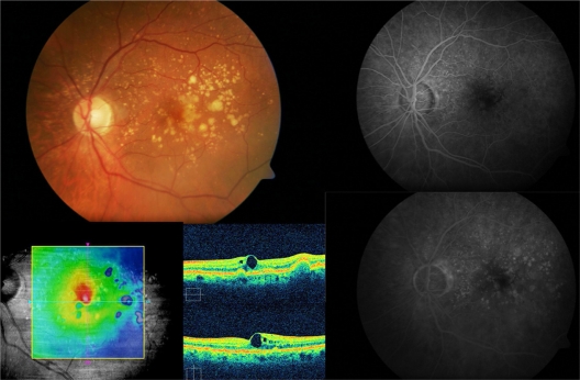
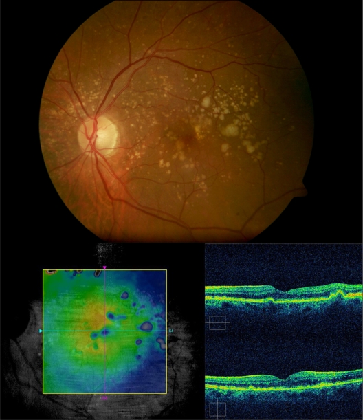
The aim of our study was to evaluate the functional and anatomic outcomes of intravitreal ranibizumab for the treatment of symptomatic drusenoid pigment epithelial detachment without choroidal neovascularization in age-related macular degeneration.
This was a prospective, single-center, uncontrolled, interventional pilot study. Six consecutive eyes (six patients) with drusenoid pigment epithelial detachment with a visual acuity of 20/63 to 20/100 and no evidence of choroidal neovascularization in age-related macular degeneration participated. Patients were given at least one intravitreal ranibizumab injection and were followed for a mean of 66.67 ± 10.3 weeks. Main outcome measures included best-corrected visual acuity (BCVA) measured by Early Treatment Diabetic Retinopathy Study charts and optical coherence tomography, and central macular thickness measured by optical coherence tomography.
The mean number of intravitreal ranibizumab injections was 3.0 at the end of follow-up. Regarding BCVA and optical coherence tomography, 33.3% of eyes gained between 19 and 21 letters of BCVA, with a median decrease in central macular thickness of 21 μm. There was a statistically significant difference between baseline and final BCVA (P = 0.046). There was a positive correlation between intraretinal fluid by optical coherence tomography and improved BCVA after intravitreal ranibizumab. Metamorphopsia disappeared completely after the first injection in all subjects, with no further recurrences. No patient developed choroidal neovascularization or atrophic changes.
Intravitreal ranibizumab demonstrated anatomic and functional benefit in patients with symptomatic drusenoid pigment epithelial detachment without choroidal neovascularization in age-related macular degeneration. Further long-term, randomized, controlled trials should be performed to confirm our preliminary results.
我们研究的目的是评估玻璃体内注射雷珠单抗治疗年龄相关性黄斑变性中无症状性玻璃膜疣样色素上皮脱离且无脉络膜新生血管形成的功能和解剖学结局。
这是一项前瞻性、单中心、非对照、干预性试点研究。纳入6例(6只眼)年龄相关性黄斑变性患者,其玻璃膜疣样色素上皮脱离,视力为20/63至20/100,且无脉络膜新生血管形成的证据。患者接受至少一次玻璃体内注射雷珠单抗,并平均随访66.67±10.3周。主要结局指标包括使用早期糖尿病视网膜病变研究图表和光学相干断层扫描测量的最佳矫正视力(BCVA),以及使用光学相干断层扫描测量的中心黄斑厚度。
随访结束时,玻璃体内注射雷珠单抗的平均次数为3.0次。关于BCVA和光学相干断层扫描,33.3%的患眼BCVA提高了19至21个字母,中心黄斑厚度中位数下降了21μm。基线和最终BCVA之间存在统计学显著差异(P = 0.046)。光学相干断层扫描显示的视网膜内液与玻璃体内注射雷珠单抗后BCVA改善之间存在正相关。所有受试者在首次注射后变形视全部消失,未再复发。无患者发生脉络膜新生血管形成或萎缩性改变。
玻璃体内注射雷珠单抗对年龄相关性黄斑变性中无症状性玻璃膜疣样色素上皮脱离且无脉络膜新生血管形成的患者具有解剖学和功能上的益处。应进行进一步的长期、随机、对照试验以证实我们的初步结果。