Unit 836, Institut National de la Santé et de la Recherche Médicale, La Tronche, Isère, France.
PLoS One. 2011 Feb 24;6(2):e17416. doi: 10.1371/journal.pone.0017416.
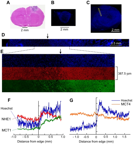
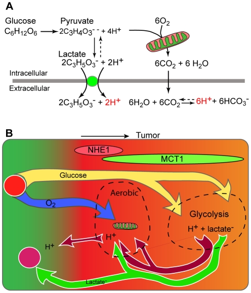
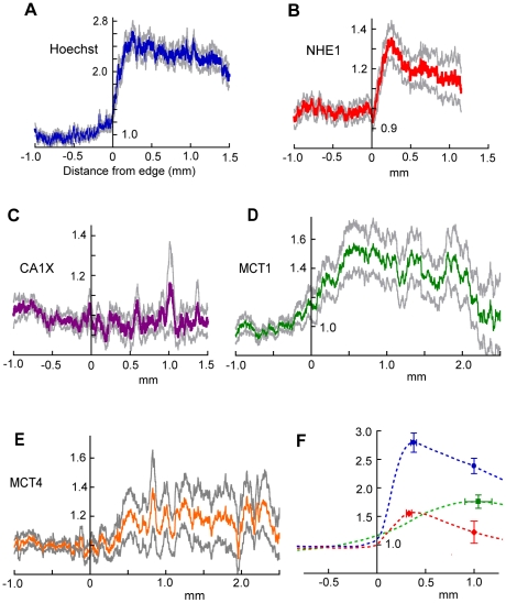
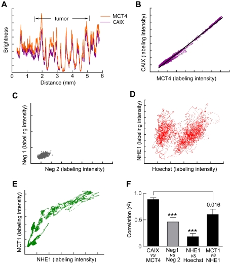
Tumors create a heterogeneous acidic microenvironment which assists their growth and which must be taken into account in the design of drugs and their delivery. In addition, the acidic extracellular pH (pHe) is itself exploited in several experimental techniques for drug delivery. The way the acidity is created is not clear. We report here the spatial organization of key proton-handling proteins in C6 gliomas in rat brain. The mean profiles across the tumor rim of the Na+/H+ exchanger NHE1, and the lactate-H+ cotransporter MCT1, both showed peaks. NHE1, which is important for extension and migration of cells in vitro, showed a peak 1.55 times higher than in extratumoural tissue at 0.33 mm from the edge. MCT1 had a broader peak, further into the tumor (maximum 1.76 fold at 1.0 mm from the edge). In contrast, MCT4 and the carbonic anhydrase CAIX, which are associated with hypoxia, were not significantly upregulated in the rim. The spatial distribution of MCT4 was highly correlated with that of CAIX, suggesting that their expression is regulated by the same factors. Since protons extruded by NHE1 diffuse away through extracellular clefts, NHE1 requires a continuous source of intracellular protons. From the stoichiometries of metabolic pathways that produce or consume H+, and the greater availability of glucose compared to oxygen in most parts of a tumor, we support the classic view that most of the net proton efflux from C6 gliomas originates in glycolytic formation of lactate and H+ inside the tumor, but add that some lactate is taken up into cells in the rim on MCT1, and some lactate diffuses away, leaving its associated protons available to re-enter cells for extrusion on NHE1. Therapeutic inhibition of NHE1, MCT1 or CAIX is predicted to affect different parts of a tumor.
肿瘤会产生一种异质的酸性微环境,这种微环境有助于肿瘤的生长,在设计药物及其输送方式时必须考虑到这一点。此外,酸性细胞外 pH 值(pHe)本身也被用于几种药物输送的实验技术中。酸度的产生方式尚不清楚。我们在此报告大鼠脑 C6 神经胶质瘤中关键质子处理蛋白的空间组织。肿瘤边缘的 Na+/H+交换器 NHE1 和乳酸-H+协同转运蛋白 MCT1 的平均分布曲线均显示出峰值。NHE1 在体外细胞的延伸和迁移中很重要,在距离边缘 0.33mm 处的峰值比肿瘤外组织高 1.55 倍。MCT1 的峰值更宽,更深入肿瘤(距离边缘 1.0mm 处的最大值为 1.76 倍)。相比之下,与缺氧相关的 MCT4 和碳酸酐酶 CAIX 在边缘处没有明显上调。MCT4 的空间分布与 CAIX 的分布高度相关,表明它们的表达受到相同因素的调控。由于 NHE1 挤出的质子通过细胞外裂隙扩散,因此 NHE1 需要不断的细胞内质子源。根据产生或消耗 H+的代谢途径的化学计量关系,以及在大多数肿瘤部位葡萄糖的可用性比氧气高,我们支持经典观点,即 C6 神经胶质瘤的大部分净质子外流源自肿瘤内的糖酵解形成乳酸和 H+,但我们补充说,一些乳酸通过 MCT1 被摄取到边缘细胞中,一些乳酸扩散出去,留下其相关的质子可重新进入细胞通过 NHE1 排出。NHE1、MCT1 或 CAIX 的治疗性抑制预计会影响肿瘤的不同部位。