Department of Surgery, Leiden University Medical Center, Leiden, The Netherlands.
J Surg Res. 2012 May 15;174(2):266-71. doi: 10.1016/j.jss.2011.01.009. Epub 2011 Feb 2.
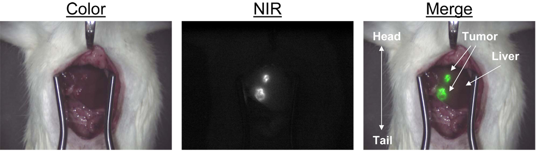
Near-infrared (NIR) fluorescence imaging using indocyanine green (ICG) is a promising technique to obtain real-time assessment of the extent and number of colorectal liver metastases during surgery. The current study aims to optimize dosage and timing of ICG administration.
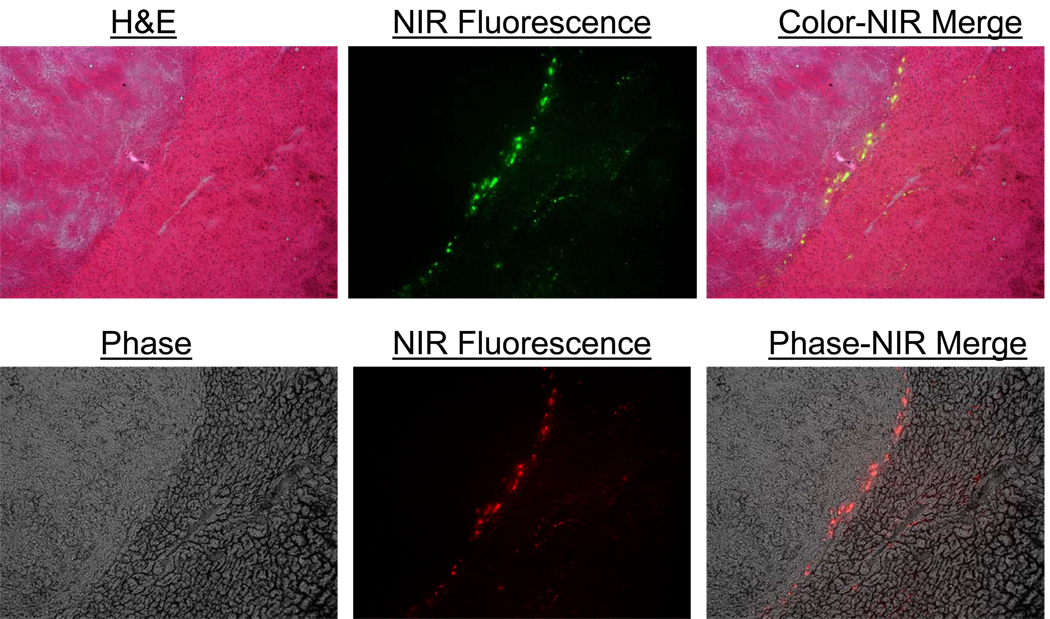
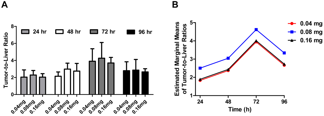
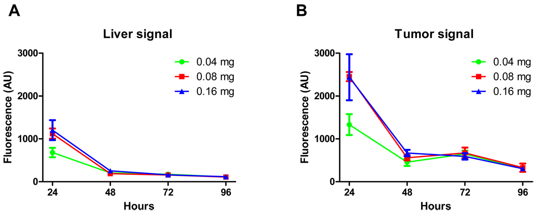
Liver tumors were induced in 18 male WAG/Rij rats by subcapsular inoculation of CC531 rat colorectal cancer cells into three distinct liver lobes. Rats were divided in two groups: imaging after 24 and 48 h or 72 and 96 h after intravenous ICG administration. In each time group, rats were allocated to three dose groups: 0.04, 0.08, or 0.16 mg ICG. Intraoperative imaging and ex vivo measurements were performed using the Mini-FLARE imaging system and confirmed by fluorescence microscopy. Fluorescence intensity was quantified using the Mini-FLARE software and the difference between tumor signal and liver signal (tumor-to-liver ratio; TLR) was calculated.
In all 18 rats, all colorectal liver metastases (n = 34), some as small as 1.2 mm, were identified using ICG and the Mini-FLARE imaging system. Average tumor-to-liver ratio (TLR) over all groups was 3.0 ± 1.2. TLR was significantly higher in the 72 h time group compared with other time points. ICG dose did not significantly influence TLR, but a trend was found favoring the 0.08 mg dose group. Fluorescence microscopy demonstrated a clear fluorescent rim around the tumor.
This study demonstrates that colorectal cancer liver metastases can be clearly identified during surgery using ICG and the Mini-FLARE imaging system, with optimal timing of 72 h post-injection and an optimal dose of 0.08 mg (0.25 mg/kg) ICG. NIR fluorescence imaging has the potential to improve intraoperative detection of micrometastases and, thus, the completeness of resection.
近红外(NIR)荧光成像是一种使用吲哚菁绿(ICG)实时评估结直肠癌肝转移手术中肝转移灶范围和数量的有前途的技术。本研究旨在优化 ICG 给药剂量和时间。
通过将 CC531 大鼠结直肠癌细胞接种到三个不同的肝叶的包膜下,在 18 只雄性 WAG/Rij 大鼠中诱导肝肿瘤。大鼠分为两组:静脉注射 ICG 后 24 小时和 48 小时或 72 小时和 96 小时进行成像。在每个时间组中,大鼠分为三组:0.04、0.08 或 0.16 mg ICG。使用 Mini-FLARE 成像系统进行术中成像和离体测量,并通过荧光显微镜进行确认。使用 Mini-FLARE 软件对荧光强度进行量化,并计算肿瘤信号与肝脏信号之间的差异(肿瘤与肝脏比值;TLR)。
在所有 18 只大鼠中,均使用 ICG 和 Mini-FLARE 成像系统识别所有结直肠肝转移灶(n = 34),其中一些小至 1.2 毫米。所有组的平均肿瘤与肝脏比值(TLR)为 3.0 ± 1.2。与其他时间点相比,72 小时时间组的 TLR 明显更高。ICG 剂量对 TLR 没有显著影响,但发现 0.08 mg 剂量组有优势。荧光显微镜显示肿瘤周围有明显的荧光环。
本研究表明,使用 ICG 和 Mini-FLARE 成像系统可以在手术中清晰地识别结直肠癌肝转移灶,最佳注射时间为注射后 72 小时,最佳剂量为 0.08 mg(0.25 mg/kg)ICG。近红外荧光成像有可能提高术中对微转移灶的检测能力,从而提高切除的完整性。