Bunyan Reem F, Popescu Bogdan F Gh, Carter Jonathan L, Caselli Richard J, Parisi Joseph E, Lucchinetti Claudia F
Department of Neurology, Mayo Clinic, College of Medicine, Rochester, MN 55905, USA.
Arch Neurol. 2011 Apr;68(4):525-8. doi: 10.1001/archneurol.2011.50.
To describe a case of childhood-onset progressive multiple sclerosis with dementia and evidence of extensive cortical demyelination from brain biopsy specimen.
Case report.
Mayo Clinic, Rochester, Minnesota.
A 26-year-old man with a history of behavioral changes starting at the age of 13 years followed by progressive dementia.
Neurological examination, magnetic resonance imaging, cerebrospinal fluid studies, neuropsychological testing, and brain biopsy.
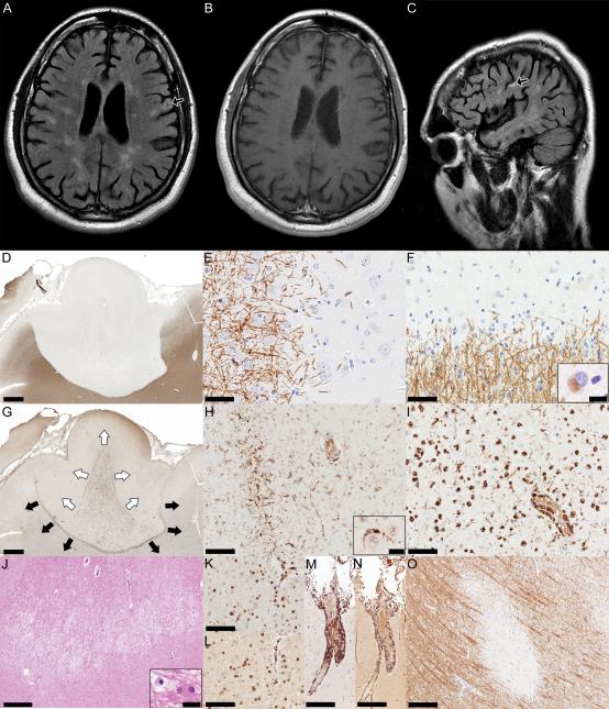
Magnetic resonance imaging scans showed numerous T2-weighted hyperintensities throughout the central nervous system not associated with contrast enhancement. Brain biopsy specimens showed cortical and subcortical demyelination. All 3 types of cortical demyelinating lesions were observed: leukocortical, intracortical, and subpial. Lesions were associated with profound microglial activation. The patient continued to progress despite attempts to treat with multiple sclerosis disease-modifying therapies.
Multiple sclerosis should be considered in the diagnosis of progressive dementia in children and young adults. Cortical demyelination may contribute to cognitive decline in patients with dementia due to multiple sclerosis.
描述一例儿童期起病的进行性多发性硬化症病例,伴有痴呆,且脑活检标本显示广泛皮质脱髓鞘证据。
病例报告。
明尼苏达州罗切斯特市梅奥诊所。
一名26岁男性,13岁开始出现行为改变,随后逐渐发展为痴呆。
神经学检查、磁共振成像、脑脊液研究、神经心理学测试和脑活检。
磁共振成像扫描显示整个中枢神经系统有大量T2加权高信号,无强化表现。脑活检标本显示皮质和皮质下脱髓鞘。观察到所有3种类型的皮质脱髓鞘病变:白质皮质、皮质内和软膜下。病变与显著的小胶质细胞激活有关。尽管尝试使用多种多发性硬化症疾病修正疗法进行治疗,患者仍继续进展。
在儿童和青年进行性痴呆的诊断中应考虑多发性硬化症。皮质脱髓鞘可能导致多发性硬化症所致痴呆患者的认知功能下降。