Digital Image Processing, Telemedicine and Multimedia Lab, Faculty of Electrical Engineering, University of Belgrade, Belgrade, Serbia.
Diagn Pathol. 2011 Mar 30;6 Suppl 1(Suppl 1):S21. doi: 10.1186/1746-1596-6-S1-S21.
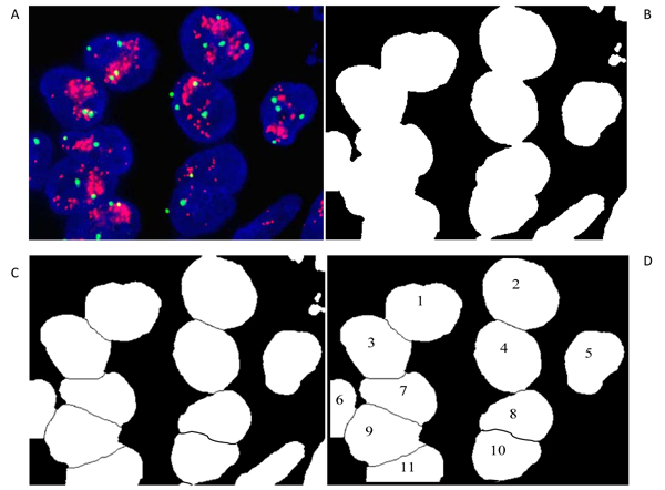
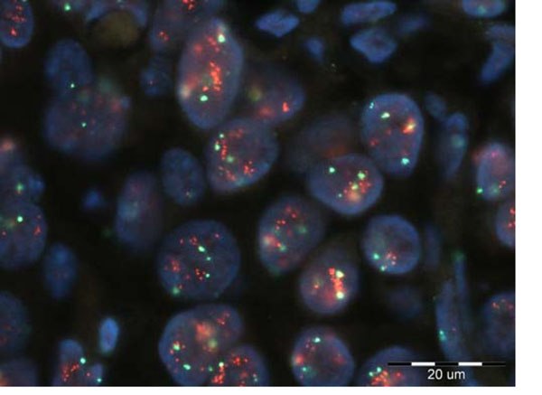
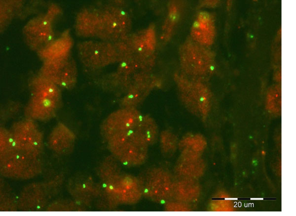
Fluorescence in situ hybridization (FISH) is very accurate method for measuring HER2 gene copies, as a sign of potential breast cancer. This method requires small tissue samples, and has a high sensitivity to detect abnormalities from a histological section. By using multiple colors, this method allows the detection of multiple targets simultaneously. The target parts in the cells become visible as colored dots. The HER-2 probes are visible as orange stained spots under a fluorescent microscope while probes for centromere 17 (CEP-17), the chromosome on which the gene HER-2/neu is located, are visible as green spots.
The conventional analysis involves the scoring of the ratio of HER-2/neu over CEP 17 dots within each cell nucleus and then averaging the scores for a number of 60 cells. A ratio of 2.0 of HER-2/neu to CEP 17 copy number denotes amplification. Several methods have been proposed for the detection and automated evaluation (dot counting) of FISH signals. In this paper the combined method based on the mathematical morphology (MM) and inverse multifractal (IMF) analysis is suggested. Similar method was applied recently in detection of microcalcifications in digital mammograms, and was very successful.
The combined MM using top-hat and bottom-hat filters, and the IMF method was applied to FISH images from Molecular Biology Lab, Department of Pathology, Wielkoposka Cancer Center, Poznan. Initial results indicate that this method can be applied to FISH images for the evaluation of HER2/neu status.
Mathematical morphology and multifractal approach are used for colored dot detection and counting in FISH images. Initial results derived on clinical cases are promising. Note that the overlapping of colored dots, particularly red/orange dots, needs additional improvements in post-processing.
荧光原位杂交(FISH)是一种非常精确的方法,可用于测量 HER2 基因拷贝数,作为潜在乳腺癌的标志。该方法需要小的组织样本,并且对从组织切片中检测异常具有很高的灵敏度。通过使用多种颜色,该方法可以同时检测多个目标。细胞中的目标部分在荧光显微镜下呈现为彩色斑点。HER-2 探针在橙色染色斑点下可见,而位于基因 HER-2/neu 所在染色体上的着丝粒 17(CEP-17)的探针则可见为绿色斑点。
传统分析包括对每个细胞核内 HER-2/neu 与 CEP 17 点的比值进行评分,然后对 60 个细胞的评分进行平均。HER-2/neu 与 CEP 17 拷贝数的比值为 2.0 表示扩增。已经提出了几种用于检测和自动评估(点计数)FISH 信号的方法。本文提出了一种基于数学形态学(MM)和逆多重分形(IMF)分析的组合方法。最近,该方法已应用于数字乳房 X 线照片中微钙化的检测,并且非常成功。
使用顶帽和底帽滤波器的组合 MM 以及 IMF 方法应用于来自病理学系分子生物学实验室的 FISH 图像,大波兰癌症中心,波兹南。初步结果表明,该方法可应用于 FISH 图像以评估 HER2/neu 状态。
数学形态学和多重分形方法用于 FISH 图像中彩色斑点的检测和计数。从临床病例中得出的初步结果很有希望。请注意,彩色斑点,特别是红色/橙色斑点的重叠,需要在后期处理中进行额外的改进。