Surgical Neurology Branch, National Institute of Neurological Disorders and Stroke, NIH, Bethesda, MD, USA.
Mod Pathol. 2011 Aug;24(8):1023-30. doi: 10.1038/modpathol.2011.61. Epub 2011 Apr 15.
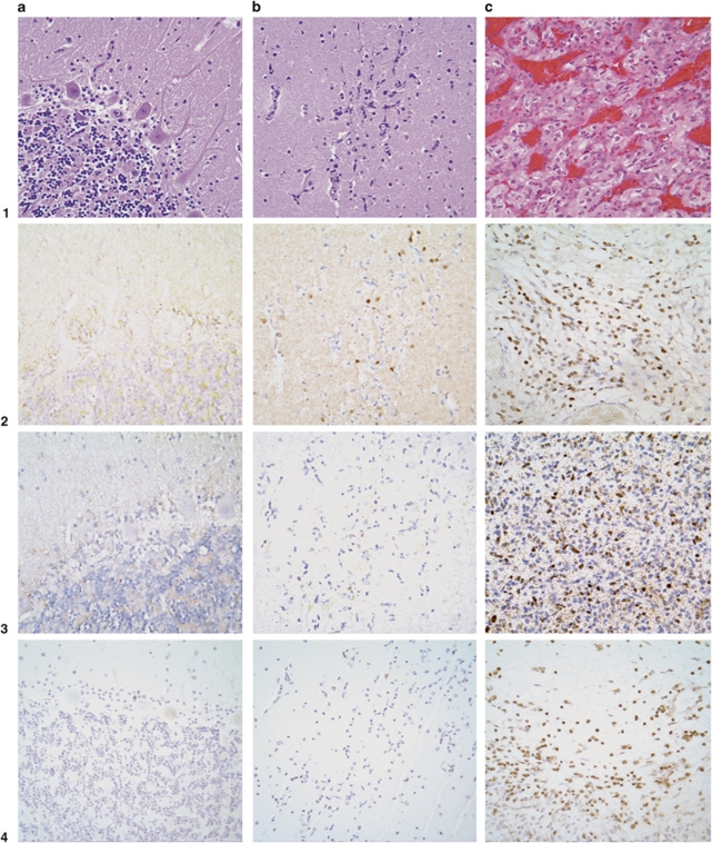
There is increasing evidence that suggests that knockout of tumor-suppressor gene function causes developmental arrest and protraction of cellular differentiation. In the peripheral nervous system of patients with the tumor-suppressor gene disorder, von Hippel-Lindau disease, we have demonstrated developmentally arrested structural elements composed of hemangioblast progenitor cells. Some developmentally arrested structural elements progress to a frank tumor, hemangioblastoma. However, in von Hippel-Lindau disease, hemangioblastomas are frequently observed in the cerebellum, suggesting an origin in the central nervous system. We performed a structural and topographic analysis of cerebellar tissues obtained from von Hippel-Lindau disease patients to identify and characterize developmentally arrested structural elements in the central nervous system. We examined the entire cerebella of five tumor-free von Hippel-Lindau disease patients and of three non-von Hippel-Lindau disease controls. In all, 9 cerebellar developmentally arrested structural elements were detected and topographically mapped in 385 blocks of von Hippel-Lindau disease cerebella. No developmentally arrested structural elements were seen in 214 blocks from control cerebella. Developmentally arrested structural elements are composed of poorly differentiated cells that express hypoxia-inducible factor (HIF)2α, but not HIF1α or brachyury, and preferentially involve the molecular layer of the dorsum cerebelli. For the first time, we identify and characterize developmentally arrested structural elements in the central nervous system of von Hippel-Lindau patients. We provide evidence that developmentally arrested structural elements in the cerebellum are composed of developmentally arrested hemangioblast progenitor cells in the molecular layer of the dorsum cerebelli.
越来越多的证据表明,肿瘤抑制基因功能的缺失会导致发育停滞和细胞分化的延长。在患有肿瘤抑制基因紊乱——von Hippel-Lindau 病的患者的周围神经系统中,我们已经证明了由成血管母细胞祖细胞组成的发育停滞的结构元素。一些发育停滞的结构元素进展为真正的肿瘤——血管母细胞瘤。然而,在 von Hippel-Lindau 病中,小脑中经常观察到血管母细胞瘤,提示其起源于中枢神经系统。我们对来自 von Hippel-Lindau 病患者的小脑组织进行了结构和拓扑分析,以鉴定和描述中枢神经系统中的发育停滞结构元素。我们检查了五名无肿瘤的 von Hippel-Lindau 病患者和三名非 von Hippel-Lindau 病对照者的整个小脑。总共在 von Hippel-Lindau 病患者的小脑的 385 个区块中检测到并绘制了 9 个发育停滞的结构元素,而在 214 个对照者小脑的区块中则未发现发育停滞的结构元素。发育停滞的结构元素由表达低氧诱导因子(HIF)2α但不表达 HIF1α或 brachyury 的分化不良细胞组成,并且优先涉及小脑背侧的分子层。我们首次在 von Hippel-Lindau 病患者的中枢神经系统中鉴定和描述了发育停滞的结构元素。我们提供的证据表明,小脑中的发育停滞结构元素由小脑背侧分子层中的发育停滞的成血管母细胞祖细胞组成。