Protein Structure Section, Macromolecular Crystallography Laboratory, National Cancer Institute, Frederick, MD 21702, USA.
J Struct Biol. 2011 Jul;175(1):73-84. doi: 10.1016/j.jsb.2011.04.009. Epub 2011 Apr 20.
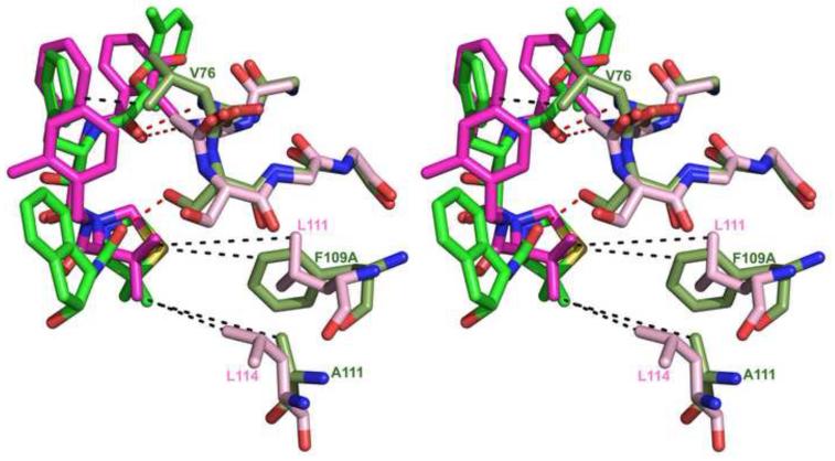
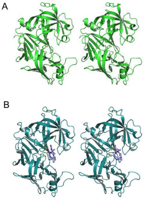
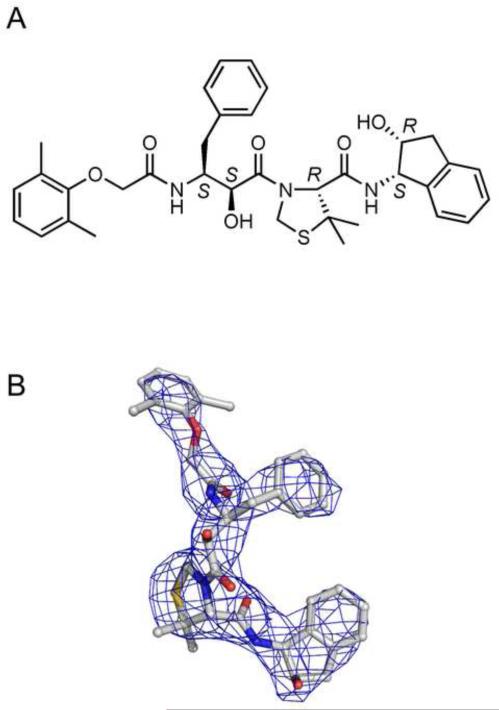
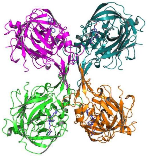
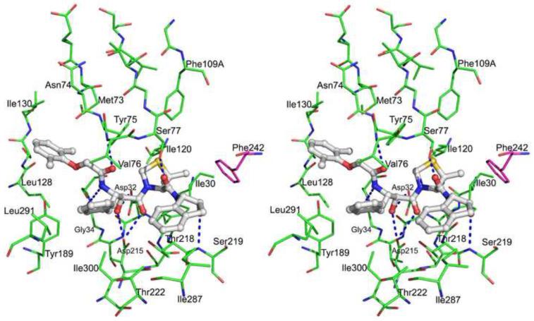
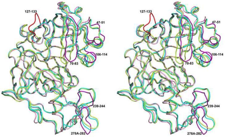
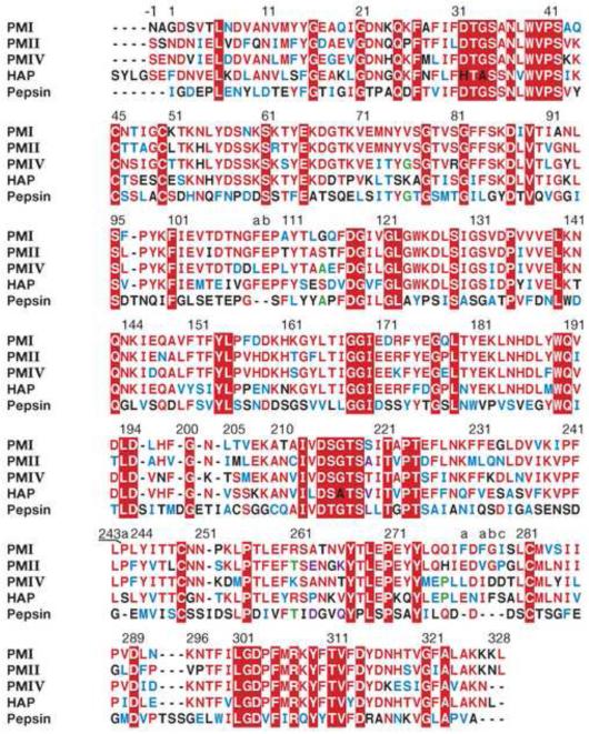
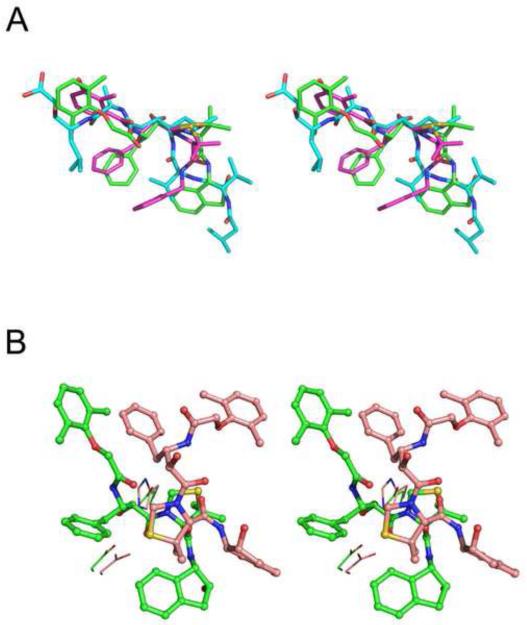
Plasmepsin I (PMI) is one of the four vacuolar pepsin-like proteases responsible for hemoglobin degradation by the malarial parasite Plasmodium falciparum, and the only one with no crystal structure reported to date. Due to substantial functional redundancy of these enzymes, lack of inhibition of even a single plasmepsin can defeat efforts in creating effective antiparasitic agents. We have now solved crystal structures of the recombinant PMI as apoenzyme and in complex with the potent peptidic inhibitor, KNI-10006, at the resolution of 2.4 and 3.1Å, respectively. The apoenzyme crystallized in the orthorhombic space group P2(1)2(1)2(1) with two molecules in the asymmetric unit and the structure has been refined to the final R-factor of 20.7%. The KNI-10006 bound enzyme crystallized in the tetragonal space group P4(3) with four molecules in the asymmetric unit and the structure has been refined to the final R-factor of 21.1%. In the PMI-KNI-10006 complex, the inhibitors were bound identically to all four enzyme molecules, with the opposite directionality of the main chain of KNI-10006 relative to the direction of the enzyme substrates. Such a mode of binding of inhibitors containing an allophenylnorstatine-dimethylthioproline insert in the P1-P1' positions, previously reported in a complex with PMIV, demonstrates the importance of satisfying the requirements for the proper positioning of the functional groups in the mechanism-based inhibitors towards the catalytic machinery of aspartic proteases, as opposed to binding driven solely by the specificity of the individual enzymes. A comparison of the structure of the PMI-KNI-10006 complex with the structures of other vacuolar plasmepsins identified the important differences between them and may help in the design of specific inhibitors targeting the individual enzymes.
疟原虫裂殖子内的 PfPMIs 是 4 种液泡型组织蛋白酶样蛋白酶之一,能降解血红蛋白,目前尚未报道其晶体结构。由于这些酶的功能存在大量冗余,缺乏对哪怕是单一 PfPMIs 的抑制都会使创建有效抗寄生虫药物的努力付诸东流。我们现已解析出重组 PfPMI 酶原和与其强肽类抑制剂 KNI-10006 复合物的晶体结构,分辨率分别为 2.4Å 和 3.1Å。酶原晶体呈正交晶系 P2(1)2(1)2(1),每不对称单位包含 2 个分子,经最终 R 因子为 20.7%的修正。KNI-10006 结合酶晶体呈四方晶系 P4(3),每不对称单位包含 4 个分子,经最终 R 因子为 21.1%的修正。在 PfPMI-KNI-10006 复合物中,抑制剂与所有 4 个酶分子的结合方式相同,抑制剂主链的方向与酶底物的方向相反。这种结合方式在与 PMIV 形成复合物时,对 P1-P1' 位置含有非天然苯丙氨酸-二甲基硫代脯氨酸插入的抑制剂也同样适用,表明了对于天冬氨酸蛋白酶的催化机制,基于机制的抑制剂中功能基团的适当定位的要求至关重要,而不仅仅是通过各个酶的特异性来驱动结合。PfPMI-KNI-10006 复合物结构与其他液泡型 PfPMIs 的结构进行比较,发现了它们之间的重要差异,这可能有助于针对个别酶设计特异性抑制剂。