Department of Microbiology and Department of Biochemistry and Molecular Biology, Monash University, Clayton, Victoria, Australia.
PLoS One. 2011 Apr 20;6(4):e18981. doi: 10.1371/journal.pone.0018981.
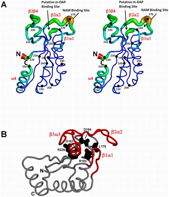
The C-terminal domain of MotB (MotB-C) shows high sequence similarity to outer membrane protein A and related peptidoglycan (PG)-binding proteins. It is believed to anchor the power-generating MotA/MotB stator unit of the bacterial flagellar motor to the peptidoglycan layer of the cell wall. We previously reported the first crystal structure of this domain and made a puzzling observation that all conserved residues that are thought to be essential for PG recognition are buried and inaccessible in the crystal structure. In this study, we tested a hypothesis that peptidoglycan binding is preceded by, or accompanied by, some structural reorganization that exposes the key conserved residues.
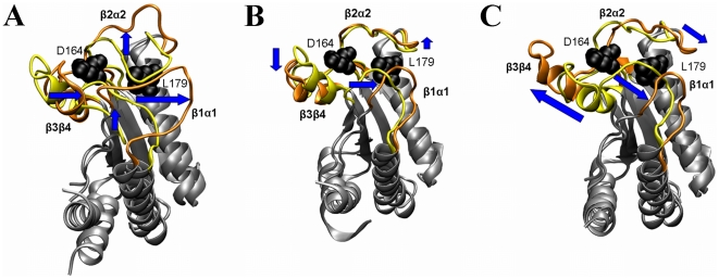
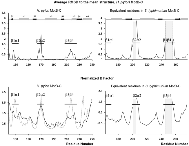
METHODOLOGY/PRINCIPAL FINDINGS: We determined the structure of a new crystalline form (Form B) of Helicobacter pylori MotB-C. Comparisons with the existing Form A revealed conformational variations in the petal-like loops around the carbohydrate binding site near one end of the β-sheet. These variations are thought to reflect natural flexibility at this site required for insertion into the peptidoglycan mesh. In order to understand the nature of this flexibility we have performed molecular dynamics simulations of the MotB-C dimer. The results are consistent with the crystallographic data and provide evidence that the three loops move in a concerted fashion, exposing conserved MotB residues that have previously been implicated in binding of the peptide moiety of peptidoglycan.
CONCLUSION/SIGNIFICANCE: Our structural analysis provides a new insight into the mechanism by which MotB inserts into the peptidoglycan mesh, thus anchoring the power-generating complex to the cell wall.
MotB 的 C 端结构域(MotB-C)与外膜蛋白 A 和相关的肽聚糖(PG)结合蛋白具有高度的序列相似性。它被认为将细菌鞭毛马达的动力产生 MotA/MotB 定子单元锚定到细胞壁的肽聚糖层上。我们之前报道了该结构域的首个晶体结构,并观察到一个令人费解的现象,即所有被认为对 PG 识别至关重要的保守残基都被埋藏且在晶体结构中无法触及。在这项研究中,我们检验了一个假设,即肽聚糖结合之前或同时发生一些结构重排,暴露出关键的保守残基。
方法/主要发现:我们确定了幽门螺杆菌 MotB-C 的一种新的晶体形式(Form B)的结构。与现有 Form A 的比较揭示了β-折叠片附近靠近碳水化合物结合位点的花瓣样环的构象变化。这些变化被认为反映了该位点自然的灵活性,这是插入肽聚糖网所必需的。为了理解这种灵活性的本质,我们对 MotB-C 二聚体进行了分子动力学模拟。结果与晶体学数据一致,并提供了证据表明,这三个环以协调的方式移动,暴露出以前被认为与肽聚糖肽部分结合相关的保守 MotB 残基。
结论/意义:我们的结构分析为 MotB 插入肽聚糖网的机制提供了新的见解,从而将动力产生复合物锚定到细胞壁上。