Department of Cell Biology, Yale University School of Medicine, New Haven, CT 06520, USA.
J Cell Biol. 2011 May 16;193(4):643-53. doi: 10.1083/jcb.201008135. Epub 2011 May 9.
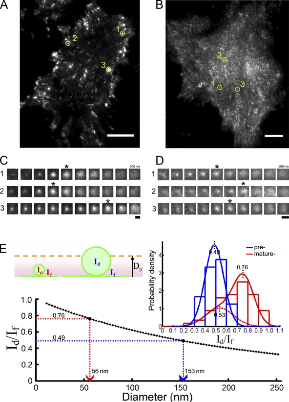
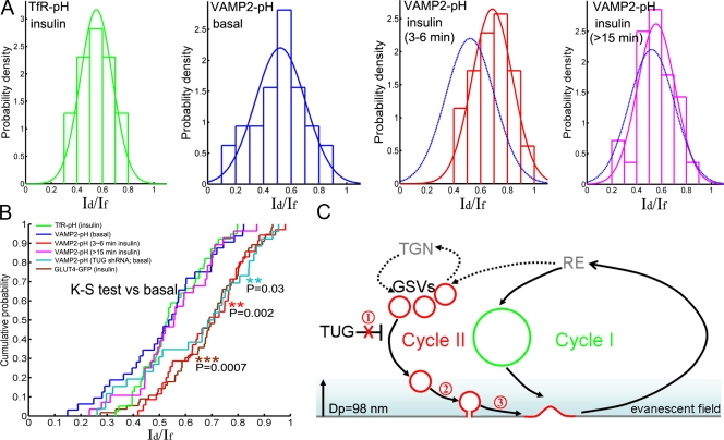
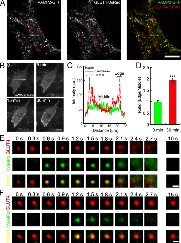
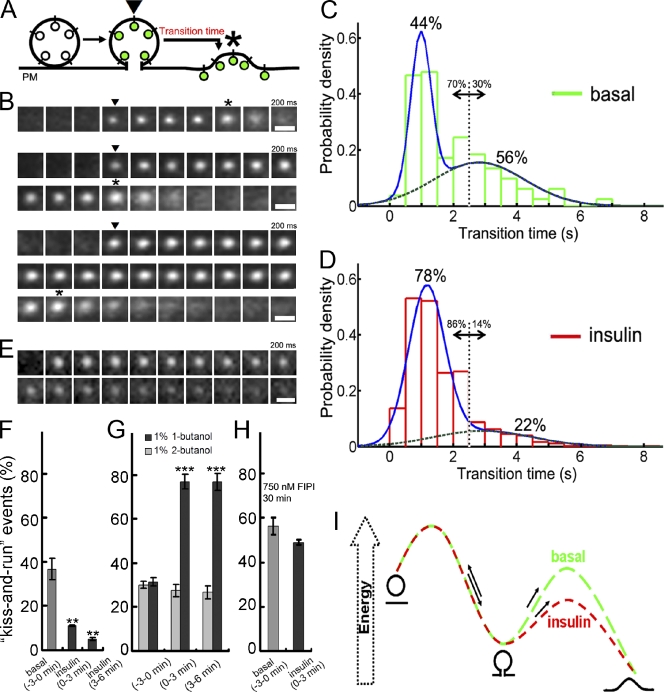
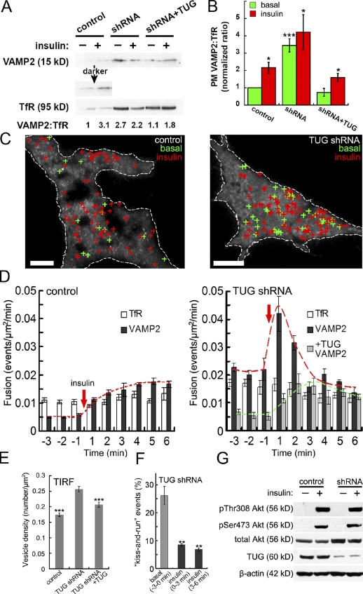
Insulin stimulates translocation of GLUT4 storage vesicles (GSVs) to the surface of adipocytes, but precisely where insulin acts is controversial. Here we quantify the size, dynamics, and frequency of single vesicle exocytosis in 3T3-L1 adipocytes. We use a new GSV reporter, VAMP2-pHluorin, and bypass insulin signaling by disrupting the GLUT4-retention protein TUG. Remarkably, in unstimulated TUG-depleted cells, the exocytic rate is similar to that in insulin-stimulated control cells. In TUG-depleted cells, insulin triggers a transient, twofold burst of exocytosis. Surprisingly, insulin promotes fusion pore expansion, blocked by acute perturbation of phospholipase D, which reflects both properties intrinsic to the mobilized vesicles and a novel regulatory site at the fusion pore itself. Prolonged stimulation causes cargo to switch from approximately 60 nm GSVs to larger exocytic vesicles characteristic of endosomes. Our results support a model whereby insulin promotes exocytic flux primarily by releasing an intracellular brake, but also by accelerating plasma membrane fusion and switching vesicle traffic between two distinct circuits.
胰岛素刺激 GLUT4 储存小泡 (GSV) 向脂肪细胞表面移位,但胰岛素的确切作用部位存在争议。在这里,我们定量分析了 3T3-L1 脂肪细胞中单囊泡胞吐的大小、动力学和频率。我们使用了一种新的 GSV 报告蛋白 VAMP2-pHluorin,并通过破坏 GLUT4 保留蛋白 TUG 来绕过胰岛素信号。值得注意的是,在未受刺激的 TUG 耗尽细胞中,胞吐率与胰岛素刺激的对照细胞相似。在 TUG 耗尽的细胞中,胰岛素触发了一个短暂的、两倍的胞吐爆发。令人惊讶的是,胰岛素促进融合孔扩张,被急性干扰磷脂酶 D 阻断,这反映了被动员的囊泡的内在特性和融合孔本身的一个新的调节位点。长时间的刺激导致货物从大约 60nm 的 GSV 切换到更大的内体特征的胞吐小泡。我们的结果支持这样一种模型,即胰岛素主要通过释放细胞内制动器来促进胞吐通量,但也通过加速质膜融合和在两个不同的回路之间切换囊泡运输来促进胞吐通量。