University of South Carolina, Program in Behavioral Neuroscience, Department of Psychology, Columbia, SC 29208, USA.
BMC Neurosci. 2011 May 10;12:38. doi: 10.1186/1471-2202-12-38.
Long-term primary neuronal cultures are a useful tool for the investigation of biochemical processes associated with neuronal senescence. Improvements in available technology make it possible to observe maturation of neural cells isolated from different regions of the rodent brain over a prolonged period in vitro. Existing experimental evidence suggests that cellular aging occurs in mature, long-term, primary neuronal cell cultures. However, detailed studies of neuronal development in vitro are needed to demonstrate the validity of long-term cell culture-based models for investigation of the biochemical mechanisms of in vitro neuronal development and senescence.
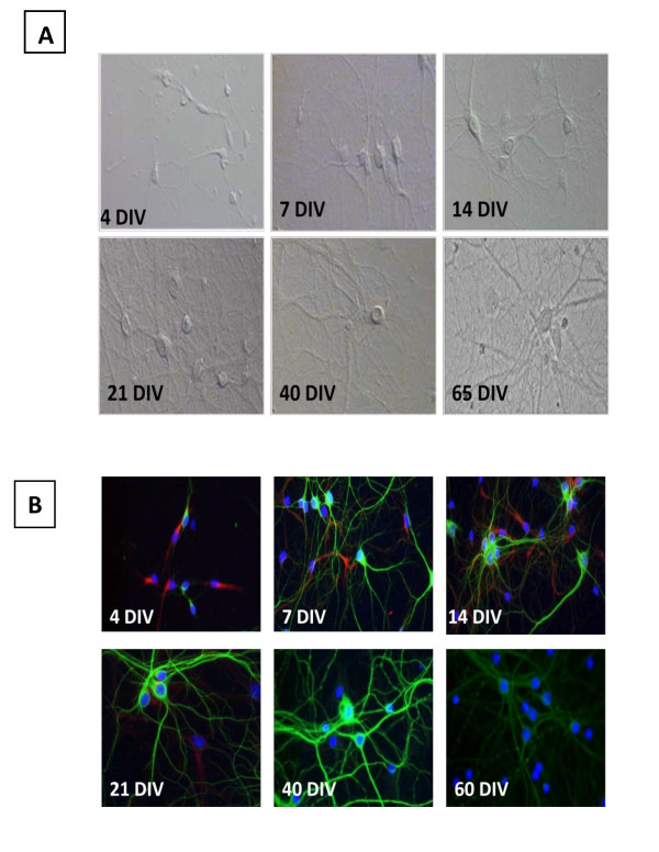
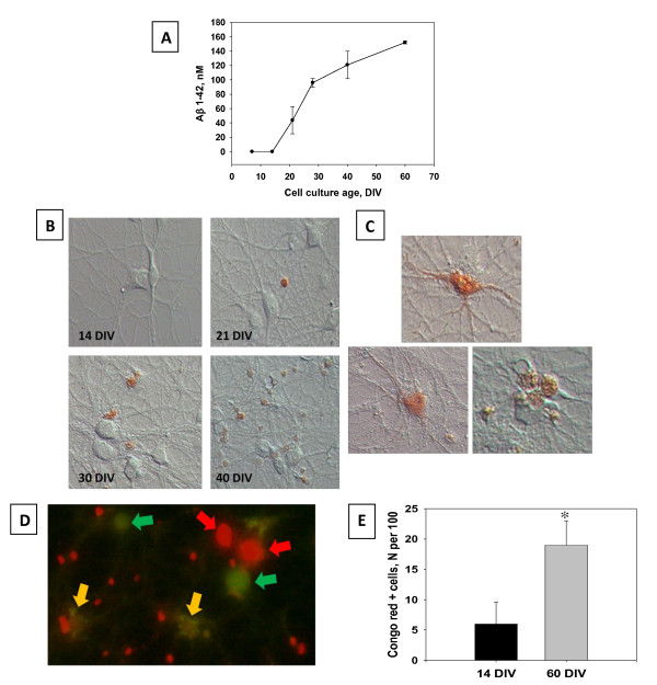
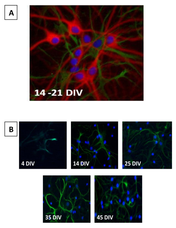
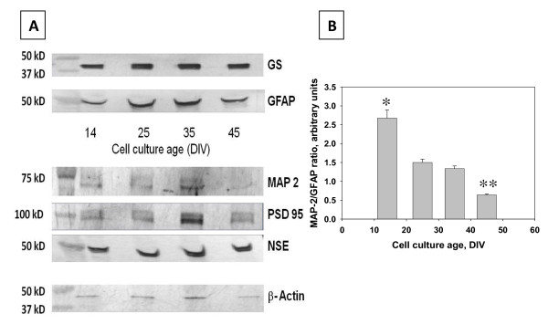
In the current study, neuron-enriched hippocampal cell cultures were used to analyze the differentiation and degeneration of hippocampal neurons over a two month time period. The expression of different neuronal and astroglial biomarkers was used to determine the cytochemical characteristics of hippocampal cells in long-term cultures of varying ages. It was observed that the expression of the intermediate filament nestin was absent from cultures older than 21 days in vitro (DIV), and the expression of neuronal or astrocytic markers appeared to replace nestin. Additionally, morphological evaluations of neuronal integrity and Hoescht staining were used to assess the cellular conditions in the process of hippocampal culture development and aging. It was found that there was an increase in endogenous production of Aβ(1-42) and an increase in the accumulation of Congo Red-binding amyloidal aggregates associated with the aging of neurons in primary culture. In vitro changes in the morphology of co-existing astrocytes and cell culture age-dependent degeneration of neurodendritic network resemble features of in vivo brain aging at the cellular level.
In conclusion, this study suggests that long-term primary CNS culture is a viable model for the study of basic mechanisms and effective methods to decelerate the process of neuronal senescence.
长期原代神经元培养是研究与神经元衰老相关的生化过程的有用工具。现有技术的改进使得有可能在体外长时间观察从啮齿动物大脑不同区域分离的神经细胞的成熟。现有的实验证据表明,细胞衰老发生在成熟的、长期的原代神经元细胞培养物中。然而,需要对体外神经发生进行详细研究,以证明长期基于细胞培养的模型用于研究体外神经发生和衰老的生化机制的有效性。
在本研究中,使用富含神经元的海马细胞培养物分析了海马神经元在两个月的时间内的分化和退化。不同神经元和星形胶质细胞生物标志物的表达用于确定不同年龄的长期培养中海马细胞的细胞化学特征。观察到在体外培养超过 21 天的培养物中不存在中间丝巢蛋白的表达,并且神经元或星形胶质细胞标志物的表达似乎取代了巢蛋白。此外,对神经元完整性的形态学评估和 Hoechst 染色用于评估海马培养物发育和老化过程中的细胞状况。发现内源性产生的 Aβ(1-42)增加,与原代培养中神经元衰老相关的刚果红结合淀粉样聚集物的积累增加。共存星形胶质细胞的体外形态变化和细胞培养年龄依赖性神经树突网络退化类似于细胞水平的体内脑衰老的特征。
总之,这项研究表明,长期中枢神经系统原代培养是研究基本机制和有效方法以减缓神经元衰老过程的可行模型。