CIBER de Enfermedades Respiratorias, Instituto de Salud Carlos III, C/ Sinesio Delgado 6, Madrid; Critical Care Center, Corporaciò Sanitaria Parc Taulí, Institut Universitari, Esfera UAB, Parc Taulí sn. Sabadell 08208; Universitat Autònoma de Barcelona, Campus de la UAB, Bellaterra 08193, Spain.
Crit Care. 2011;15(3):R124. doi: 10.1186/cc10230. Epub 2011 May 13.
Survivors of critical illness often have significant long-term brain dysfunction, and routine clinical procedures like mechanical ventilation (MV) may affect long-term brain outcome. We aimed to investigate the effect of the increase of tidal volume (Vt) on brain activation in a rat model.
Male Sprague Dawley rats were randomized to three groups: 1) Basal: anesthetized unventilated animals, 2) low Vt (LVt): MV for three hours with Vt 8 ml/kg and zero positive end-expiratory pressure (ZEEP), and 3) high Vt (HVt) MV for three hours with Vt 30 ml/kg and ZEEP. We measured lung mechanics, mean arterial pressure (MAP), arterial blood gases, and plasma and lung levels of cytokines. We used immunohistochemistry to examine c-fos as a marker of neuronal activation. An additional group of spontaneously breathing rats was added to discriminate the effect of surgical procedure and anesthesia in the brain.
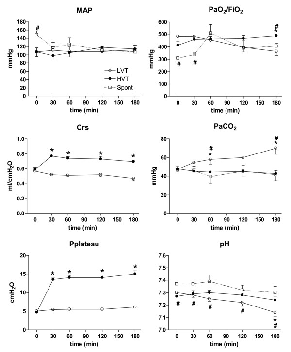
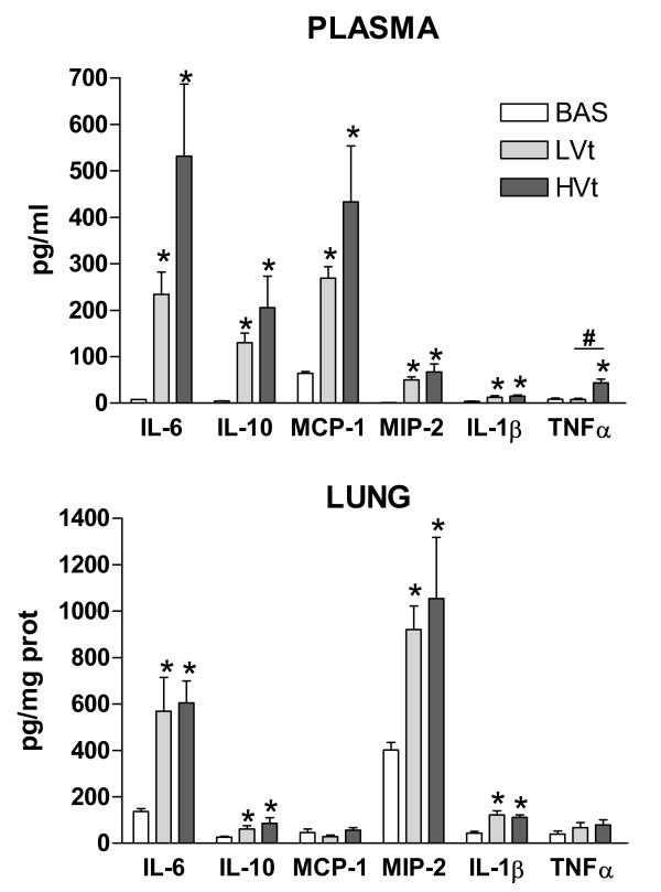
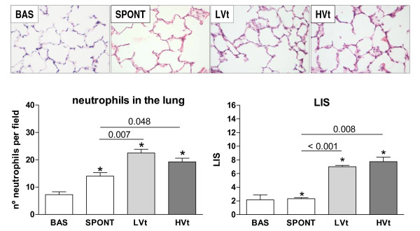
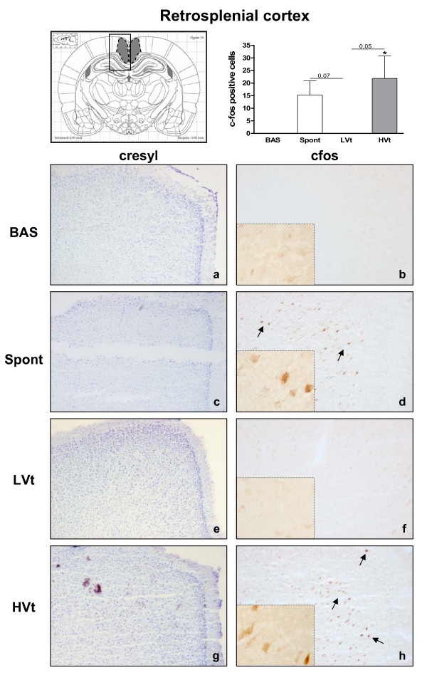
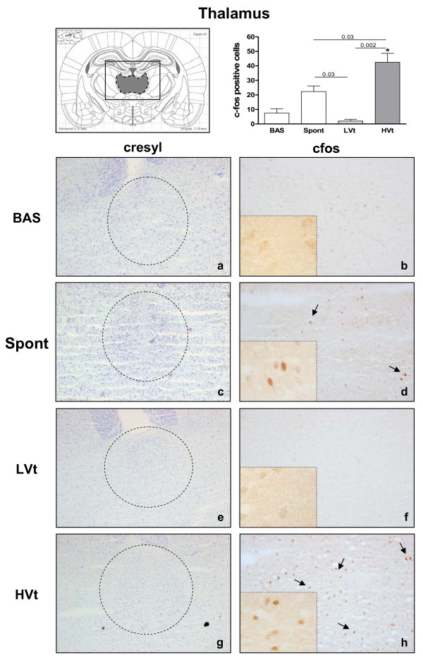
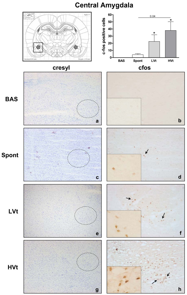
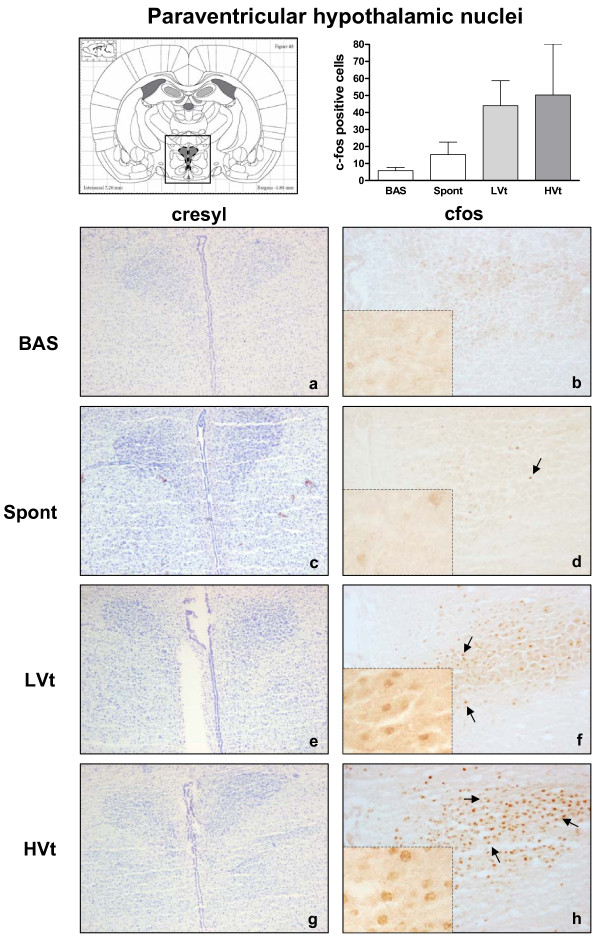
After three hours on LVt, PaO2 decreased and PaCO2 increased significantly. MAP and compliance remained stable in MV groups. Systemic and pulmonary inflammation was higher in MV rats than in unventilated rats. Plasma TNFα was significantly higher in HVt than in LVt. Immunopositive cells to c-fos in the retrosplenial cortex and thalamus increased significantly in HVt rats but not in LVt or unventilated rats.
MV promoted brain activation. The intensity of the response was higher in HVt animals, suggesting an iatrogenic effect of MV on the brain. These findings suggest that this novel cross-talking mechanism between the lung and the brain should be explored in patients undergoing MV.
危重病幸存者常存在严重的长期脑功能障碍,而机械通气(MV)等常规临床操作可能会影响长期脑结局。我们旨在研究潮气量(Vt)增加对大鼠模型脑激活的影响。
雄性 Sprague Dawley 大鼠随机分为三组:1)基础组:麻醉未通气动物,2)低 Vt 组(LVt):MV 通气 3 小时,Vt 8ml/kg,无呼气末正压(ZEEP),3)高 Vt 组(HVt):MV 通气 3 小时,Vt 30ml/kg,ZEEP。我们测量肺力学、平均动脉压(MAP)、动脉血气以及血浆和肺组织细胞因子水平。我们使用免疫组化法检测 c-fos 作为神经元激活的标志物。增加一组自主呼吸大鼠以区分手术和麻醉对大脑的影响。
LVt 通气 3 小时后,PaO2 显著降低,PaCO2 显著升高。MV 组的 MAP 和顺应性保持稳定。MV 大鼠的全身和肺部炎症较未通气大鼠明显升高。HVt 组的血浆 TNFα 明显高于 LVt 组。HVt 大鼠的后扣带回皮层和丘脑的 c-fos 免疫阳性细胞显著增加,但 LVt 或未通气大鼠则没有。
MV 促进了大脑激活。HVt 动物的反应强度更高,提示 MV 对大脑具有医源性作用。这些发现表明,应在接受 MV 的患者中探索这种肺与脑之间的新型串扰机制。