• 文献检索
  • 文档翻译
  • 深度研究
  • 学术资讯
  • Suppr Zotero 插件Zotero 插件
  • 邀请有礼
  • 套餐&价格
  • 历史记录
应用&插件
Suppr Zotero 插件Zotero 插件浏览器插件Mac 客户端Windows 客户端微信小程序
定价
高级版会员购买积分包购买API积分包
服务
文献检索文档翻译深度研究API 文档MCP 服务
关于我们
关于 Suppr公司介绍联系我们用户协议隐私条款
关注我们

Suppr 超能文献

核心技术专利:CN118964589B侵权必究
粤ICP备2023148730 号-1Suppr @ 2026

文献检索

告别复杂PubMed语法,用中文像聊天一样搜索,搜遍4000万医学文献。AI智能推荐,让科研检索更轻松。

立即免费搜索

文件翻译

保留排版,准确专业,支持PDF/Word/PPT等文件格式,支持 12+语言互译。

免费翻译文档

深度研究

AI帮你快速写综述,25分钟生成高质量综述,智能提取关键信息,辅助科研写作。

立即免费体验

黏附钙黏蛋白的黏附二聚体β链交换的分子设计原理。

Molecular design principles underlying β-strand swapping in the adhesive dimerization of cadherins.

机构信息

Department of Biochemistry and Molecular Biophysics, Columbia University, New York, New York, USA. Center for Computational Biology and Bioinformatics, Columbia University, New York, New York, USA. Howard Hughes Medical Institute, Columbia University, New York, New York, USA.

出版信息

Nat Struct Mol Biol. 2011 Jun;18(6):693-700. doi: 10.1038/nsmb.2051. Epub 2011 May 15.

DOI:10.1038/nsmb.2051
PMID:21572446
原文链接:https://pmc.ncbi.nlm.nih.gov/articles/PMC3113550/
Abstract

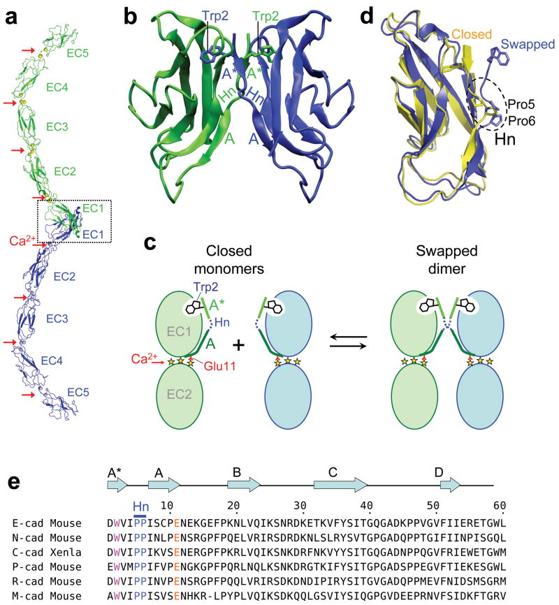
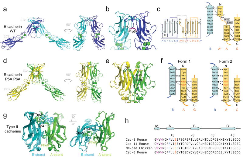
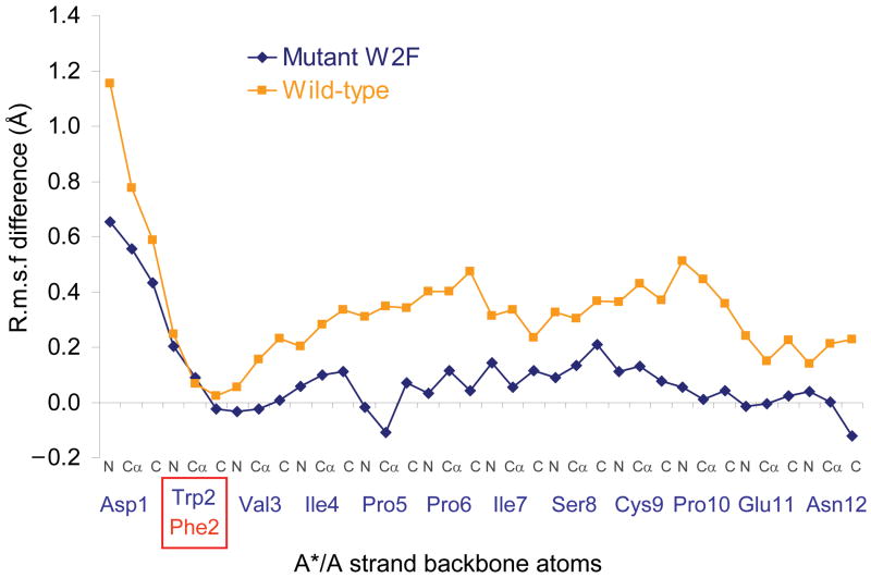
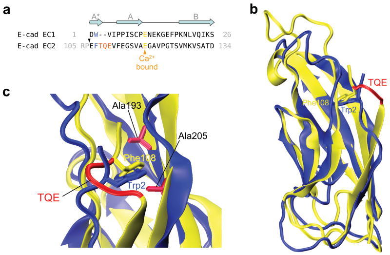
Cell adhesion by classical cadherins is mediated by dimerization of their EC1 domains through the 'swapping' of N-terminal β-strands. We use molecular simulations, measurements of binding affinities and X-ray crystallography to provide a detailed picture of the structural and energetic factors that control the adhesive dimerization of cadherins. We show that strand swapping in EC1 is driven by conformational strain in cadherin monomers that arises from the anchoring of their short N-terminal strand at one end by the conserved Trp2 and at the other by ligation to Ca(2+) ions. We also demonstrate that a conserved proline-proline motif functions to avoid the formation of an overly tight interface where affinity differences between different cadherins, crucial at the cellular level, are lost. We use these findings to design site-directed mutations that transform a monomeric EC2-EC3 domain cadherin construct into a strand-swapped dimer.

摘要

经典钙黏蛋白通过 EC1 结构域二聚化介导细胞黏附,其通过“交换”N 端β-链实现。我们使用分子模拟、结合亲和力测量和 X 射线晶体学,为控制钙黏蛋白黏附二聚化的结构和能量因素提供了详细的描述。我们表明,EC1 中的链交换是由钙黏蛋白单体中的构象应变驱动的,这种构象应变源于其短 N 端链在一端由保守的色氨酸 2 固定,另一端与 Ca(2+) 离子连接。我们还证明,一个保守的脯氨酸-脯氨酸基序可避免形成过于紧密的界面,而不同钙黏蛋白之间的亲和力差异在细胞水平上是至关重要的,在这种界面中会丧失亲和力差异。我们利用这些发现设计了定点突变,将单体 EC2-EC3 结构域钙黏蛋白构建体转化为链交换二聚体。

https://cdn.ncbi.nlm.nih.gov/pmc/blobs/119c/3113550/bf69ed69c973/nihms276132f6.jpg
https://cdn.ncbi.nlm.nih.gov/pmc/blobs/119c/3113550/86b63842f05a/nihms276132f1.jpg
https://cdn.ncbi.nlm.nih.gov/pmc/blobs/119c/3113550/f7961fa5b508/nihms276132f2.jpg
https://cdn.ncbi.nlm.nih.gov/pmc/blobs/119c/3113550/4dcdad92ce12/nihms276132f3.jpg
https://cdn.ncbi.nlm.nih.gov/pmc/blobs/119c/3113550/88b50f22cbe9/nihms276132f4.jpg
https://cdn.ncbi.nlm.nih.gov/pmc/blobs/119c/3113550/b17940d4a579/nihms276132f5.jpg
https://cdn.ncbi.nlm.nih.gov/pmc/blobs/119c/3113550/bf69ed69c973/nihms276132f6.jpg

相似文献

1
Molecular design principles underlying β-strand swapping in the adhesive dimerization of cadherins.黏附钙黏蛋白的黏附二聚体β链交换的分子设计原理。
Nat Struct Mol Biol. 2011 Jun;18(6):693-700. doi: 10.1038/nsmb.2051. Epub 2011 May 15.
2
Sequence and structural determinants of strand swapping in cadherin domains: do all cadherins bind through the same adhesive interface?钙黏蛋白结构域中链交换的序列和结构决定因素:所有钙黏蛋白都通过相同的黏附界面结合吗?
J Mol Biol. 2008 May 9;378(4):954-68. doi: 10.1016/j.jmb.2008.02.063. Epub 2008 Mar 4.
3
Mechanism of E-cadherin dimerization probed by NMR relaxation dispersion.通过 NMR 弛豫弥散研究 E-钙黏蛋白二聚体的形成机制。
Proc Natl Acad Sci U S A. 2013 Oct 8;110(41):16462-7. doi: 10.1073/pnas.1314303110. Epub 2013 Sep 25.
4
Two-step adhesive binding by classical cadherins.经典钙黏蛋白的两步黏附结合。
Nat Struct Mol Biol. 2010 Mar;17(3):348-57. doi: 10.1038/nsmb.1784. Epub 2010 Feb 28.
5
Dimerization of Cadherin-11 involves multi-site coupled unfolding and strand swapping.钙黏蛋白-11的二聚化涉及多位点耦合去折叠和链交换。
Structure. 2021 Oct 7;29(10):1105-1115.e6. doi: 10.1016/j.str.2021.06.006. Epub 2021 Jun 23.
6
Crystal structures of Drosophila N-cadherin ectodomain regions reveal a widely used class of Ca²+-free interdomain linkers.果蝇 N-钙黏蛋白外域结构域的晶体结构揭示了一类广泛使用的 Ca²⁺游离的结构域间连接体。
Proc Natl Acad Sci U S A. 2012 Jan 17;109(3):E127-34. doi: 10.1073/pnas.1117538108. Epub 2011 Dec 14.
7
Specificity of cell-cell adhesion by classical cadherins: Critical role for low-affinity dimerization through beta-strand swapping.经典钙黏蛋白介导的细胞间黏附特异性:通过β链交换形成低亲和力二聚体的关键作用。
Proc Natl Acad Sci U S A. 2005 Jun 14;102(24):8531-6. doi: 10.1073/pnas.0503319102. Epub 2005 Jun 3.
8
T-cadherin structures reveal a novel adhesive binding mechanism.T 钙黏蛋白结构揭示了一种新颖的黏附结合机制。
Nat Struct Mol Biol. 2010 Mar;17(3):339-47. doi: 10.1038/nsmb.1781. Epub 2010 Feb 28.
9
Characterizing the initial encounter complex in cadherin adhesion.描述钙黏蛋白黏附的初始接触复合物。
Structure. 2009 Aug 12;17(8):1075-81. doi: 10.1016/j.str.2009.06.012. Epub 2009 Jul 30.
10
Dynamic properties of a type II cadherin adhesive domain: implications for the mechanism of strand-swapping of classical cadherins.II型钙黏蛋白黏附结构域的动态特性:对经典钙黏蛋白链交换机制的启示
Structure. 2008 Aug 6;16(8):1195-205. doi: 10.1016/j.str.2008.05.009.

引用本文的文献

1
Molecular exaptation by the integrin αI domain.整合素αI结构域的分子扩展适应
Sci Adv. 2025 Sep 12;11(37):eadx9567. doi: 10.1126/sciadv.adx9567. Epub 2025 Sep 10.
2
IgStrand: A universal residue numbering scheme for the immunoglobulin-fold (Ig-fold) to study Ig-proteomes and Ig-interactomes.IgStrand:一种用于免疫球蛋白折叠(Ig折叠)的通用残基编号方案,用于研究Ig蛋白质组和Ig相互作用组。
PLoS Comput Biol. 2025 Apr 14;21(4):e1012813. doi: 10.1371/journal.pcbi.1012813. eCollection 2025 Apr.
3
Ig or Not Ig? That Is the Question: The Nucleating Supersecondary Structure of the Ig-Fold and the Extended Ig Universe.

本文引用的文献

1
Processing of X-ray diffraction data collected in oscillation mode.振荡模式下收集的X射线衍射数据的处理。
Methods Enzymol. 1997;276:307-26. doi: 10.1016/S0076-6879(97)76066-X.
2
The extracellular architecture of adherens junctions revealed by crystal structures of type I cadherins.I 型钙黏蛋白晶体结构揭示黏着连接的细胞外结构。
Structure. 2011 Feb 9;19(2):244-56. doi: 10.1016/j.str.2010.11.016.
3
Features and development of Coot.Coot的特点与发展
Ig 还是非 Ig?这是个问题:Ig 折叠的成核超二级结构和扩展的 Ig 宇宙。
Methods Mol Biol. 2025;2870:371-396. doi: 10.1007/978-1-0716-4213-9_19.
4
Cardiac desmosomal adhesion relies on ideal-, slip- and catch bonds.心脏桥粒黏附依赖理想黏附、滑动黏附和捕获黏附。
Sci Rep. 2024 Jan 31;14(1):2555. doi: 10.1038/s41598-024-52725-w.
5
Strengthening E-cadherin adhesion via antibody-mediated binding stabilization.通过抗体介导的结合稳定作用增强 E-钙黏蛋白黏附。
Structure. 2024 Feb 1;32(2):217-227.e3. doi: 10.1016/j.str.2023.11.002. Epub 2023 Dec 4.
6
Engineering the Interactions of Classical Cadherin Cell-Cell Adhesion Proteins.工程化经典钙黏蛋白细胞-细胞黏附蛋白的相互作用。
J Immunol. 2023 Aug 1;211(3):343-349. doi: 10.4049/jimmunol.2300098.
7
Antiparallel dimer structure of CELSR cadherin in solution revealed by high-speed atomic force microscopy.高速原子力显微镜揭示了 CELSR 钙黏蛋白在溶液中的反平行二聚体结构。
Proc Natl Acad Sci U S A. 2023 May 2;120(18):e2302047120. doi: 10.1073/pnas.2302047120. Epub 2023 Apr 24.
8
Regulation of multiple dimeric states of E-cadherin by adhesion activating antibodies revealed through Cryo-EM and X-ray crystallography.通过冷冻电镜和X射线晶体学揭示黏附激活抗体对E-钙黏蛋白多种二聚体状态的调控。
PNAS Nexus. 2022 Aug 19;1(4):pgac163. doi: 10.1093/pnasnexus/pgac163. eCollection 2022 Sep.
9
Molecular mechanism for strengthening E-cadherin adhesion using a monoclonal antibody.使用单克隆抗体增强 E-钙黏蛋白黏附的分子机制。
Proc Natl Acad Sci U S A. 2022 Aug 9;119(32):e2204473119. doi: 10.1073/pnas.2204473119. Epub 2022 Aug 3.
10
Multiple dimeric structures and strand-swap dimerization of E-cadherin in solution visualized by high-speed atomic force microscopy.高速原子力显微镜可视化的 E-钙黏蛋白在溶液中的多二聚体结构和链交换二聚化。
Proc Natl Acad Sci U S A. 2022 Jul 26;119(30):e2208067119. doi: 10.1073/pnas.2208067119. Epub 2022 Jul 22.
Acta Crystallogr D Biol Crystallogr. 2010 Apr;66(Pt 4):486-501. doi: 10.1107/S0907444910007493. Epub 2010 Mar 24.
4
T-cadherin structures reveal a novel adhesive binding mechanism.T 钙黏蛋白结构揭示了一种新颖的黏附结合机制。
Nat Struct Mol Biol. 2010 Mar;17(3):339-47. doi: 10.1038/nsmb.1781. Epub 2010 Feb 28.
5
Two-step adhesive binding by classical cadherins.经典钙黏蛋白的两步黏附结合。
Nat Struct Mol Biol. 2010 Mar;17(3):348-57. doi: 10.1038/nsmb.1784. Epub 2010 Feb 28.
6
Structure and biochemistry of cadherins and catenins.钙黏蛋白和连环蛋白的结构与生物化学。
Cold Spring Harb Perspect Biol. 2009 Sep;1(3):a003053. doi: 10.1101/cshperspect.a003053.
7
Molecular replacement with MOLREP.使用MOLREP进行分子置换。
Acta Crystallogr D Biol Crystallogr. 2010 Jan;66(Pt 1):22-5. doi: 10.1107/S0907444909042589. Epub 2009 Dec 21.
8
MolProbity: all-atom structure validation for macromolecular crystallography.MolProbity:用于大分子晶体学的全原子结构验证
Acta Crystallogr D Biol Crystallogr. 2010 Jan;66(Pt 1):12-21. doi: 10.1107/S0907444909042073. Epub 2009 Dec 21.
9
Characterizing the initial encounter complex in cadherin adhesion.描述钙黏蛋白黏附的初始接触复合物。
Structure. 2009 Aug 12;17(8):1075-81. doi: 10.1016/j.str.2009.06.012. Epub 2009 Jul 30.
10
Linking molecular affinity and cellular specificity in cadherin-mediated adhesion.在钙黏蛋白介导的黏附中连接分子亲和力与细胞特异性
Proc Natl Acad Sci U S A. 2009 Jul 14;106(28):11594-9. doi: 10.1073/pnas.0905349106. Epub 2009 Jun 24.