• 文献检索
  • 文档翻译
  • 深度研究
  • 学术资讯
  • Suppr Zotero 插件Zotero 插件
  • 邀请有礼
  • 套餐&价格
  • 历史记录
应用&插件
Suppr Zotero 插件Zotero 插件浏览器插件Mac 客户端Windows 客户端微信小程序
定价
高级版会员购买积分包购买API积分包
服务
文献检索文档翻译深度研究API 文档MCP 服务
关于我们
关于 Suppr公司介绍联系我们用户协议隐私条款
关注我们

Suppr 超能文献

核心技术专利:CN118964589B侵权必究
粤ICP备2023148730 号-1Suppr @ 2026

文献检索

告别复杂PubMed语法,用中文像聊天一样搜索,搜遍4000万医学文献。AI智能推荐,让科研检索更轻松。

立即免费搜索

文件翻译

保留排版,准确专业,支持PDF/Word/PPT等文件格式,支持 12+语言互译。

免费翻译文档

深度研究

AI帮你快速写综述,25分钟生成高质量综述,智能提取关键信息,辅助科研写作。

立即免费体验

快速进展性 MDV 驱动的 CD4+ 淋巴瘤和反应性 CD8+ T 细胞的克隆结构。

Clonal structure of rapid-onset MDV-driven CD4+ lymphomas and responding CD8+ T cells.

机构信息

Avian Infectious Disease Programme, Institute for Animal Health, Compton, Berkshire, United Kingdom.

出版信息

PLoS Pathog. 2011 May;7(5):e1001337. doi: 10.1371/journal.ppat.1001337. Epub 2011 May 5.

DOI:10.1371/journal.ppat.1001337
PMID:21573129
原文链接:https://pmc.ncbi.nlm.nih.gov/articles/PMC3088711/
Abstract

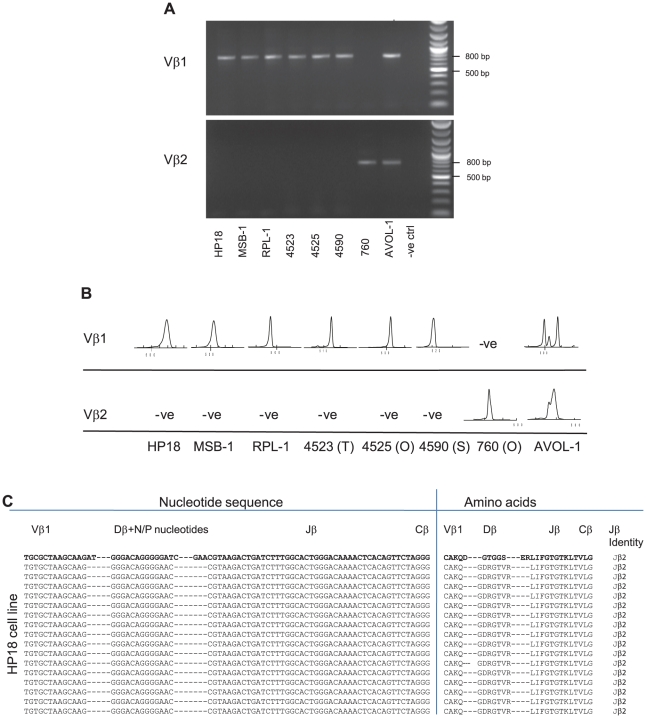
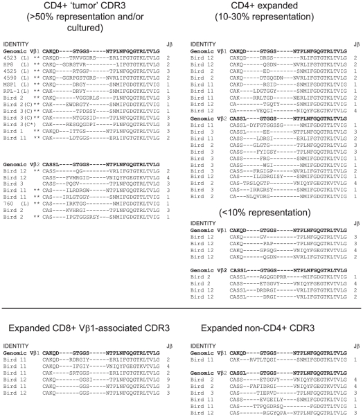
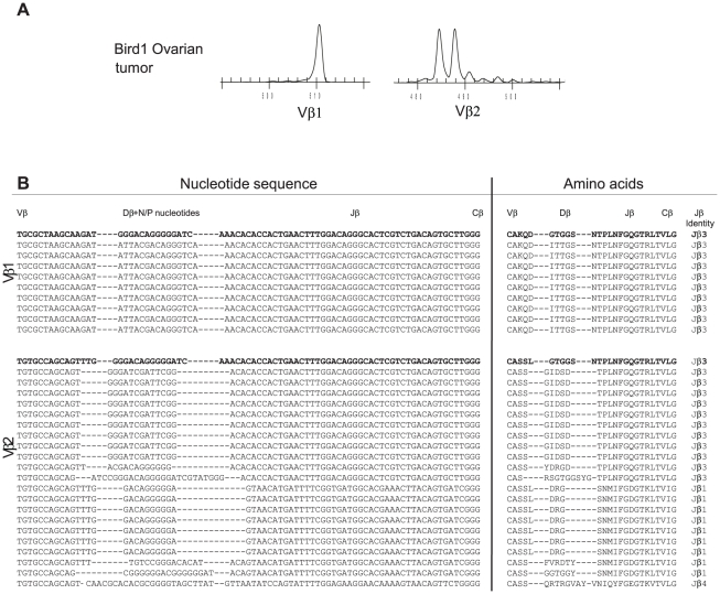
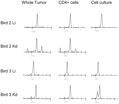
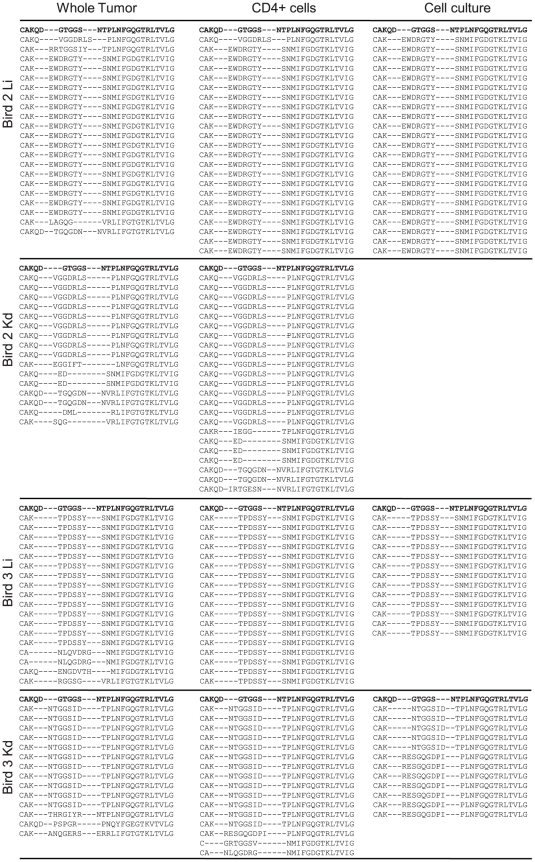
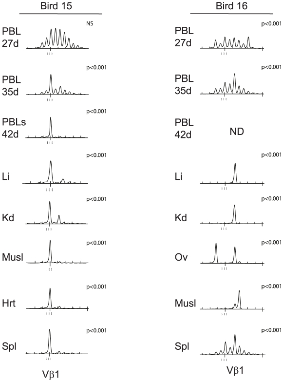
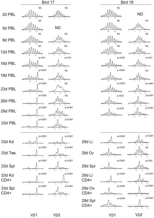
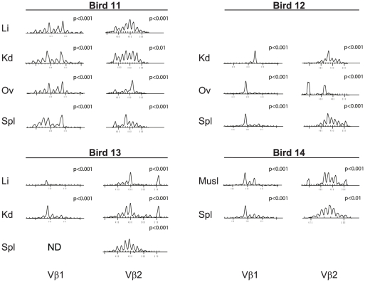
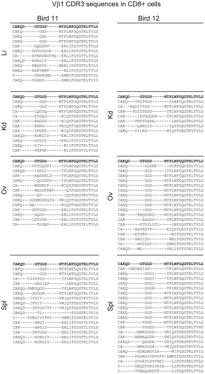
Lymphoid oncogenesis is a life threatening complication associated with a number of persistent viral infections (e.g. EBV and HTLV-1 in humans). With many of these infections it is difficult to study their natural history and the dynamics of tumor formation. Marek's Disease Virus (MDV) is a prevalent α-herpesvirus of poultry, inducing CD4+ TCRαβ+ T cell tumors in susceptible hosts. The high penetrance and temporal predictability of tumor induction raises issues related to the clonal structure of these lymphomas. Similarly, the clonality of responding CD8 T cells that infiltrate the tumor sites is unknown. Using TCRβ repertoire analysis tools, we demonstrated that MDV driven CD4+ T cell tumors were dominated by one to three large clones within an oligoclonal framework of smaller clones of CD4+ T cells. Individual birds had multiple tumor sites, some the result of metastasis (i.e. shared dominant clones) and others derived from distinct clones of transformed cells. The smaller oligoclonal CD4+ cells may represent an anti-tumor response, although on one occasion a low frequency clone was transformed and expanded after culture. Metastatic tumor clones were detected in the blood early during infection and dominated the circulating T cell repertoire, leading to MDV associated immune suppression. We also demonstrated that the tumor-infiltrating CD8+ T cell response was dominated by large oligoclonal expansions containing both "public" and "private" CDR3 sequences. The frequency of CD8+ T cell CDR3 sequences suggests initial stimulation during the early phases of infection. Collectively, our results indicate that MDV driven tumors are dominated by a highly restricted number of CD4+ clones. Moreover, the responding CD8+ T cell infiltrate is oligoclonal indicating recognition of a limited number of MDV antigens. These studies improve our understanding of the biology of MDV, an important poultry pathogen and a natural infection model of virus-induced tumor formation.

摘要

淋巴肿瘤发生是一种危及生命的并发症,与多种持续性病毒感染有关(例如人类中的 EBV 和 HTLV-1)。对于许多这些感染,很难研究其自然史和肿瘤形成的动态。马立克氏病病毒(MDV)是一种普遍存在的α疱疹病毒,可在易感宿主中诱导 CD4+TCRαβ+T 细胞肿瘤。肿瘤诱导的高穿透率和时间可预测性引发了与这些淋巴瘤克隆结构相关的问题。同样,浸润肿瘤部位的反应性 CD8+T 细胞的克隆性也未知。使用 TCRβ 库分析工具,我们证明 MDV 驱动的 CD4+T 细胞肿瘤由一个至三个大克隆主导,这些克隆在较小的 CD4+T 细胞克隆的寡克隆框架内。个别鸟类有多个肿瘤部位,有些是转移的结果(即共享主导克隆),而另一些则来自转化细胞的不同克隆。较小的寡克隆 CD4+细胞可能代表抗肿瘤反应,尽管有一次低频克隆在培养后被转化和扩增。在感染早期就可以在血液中检测到转移性肿瘤克隆,并主导循环 T 细胞库,导致与 MDV 相关的免疫抑制。我们还证明,肿瘤浸润的 CD8+T 细胞反应由包含“公共”和“私有”CDR3 序列的大寡克隆扩增主导。CD8+T 细胞 CDR3 序列的频率表明在感染的早期阶段进行了初始刺激。总的来说,我们的研究结果表明,MDV 驱动的肿瘤主要由高度受限数量的 CD4+克隆主导。此外,反应性 CD8+T 细胞浸润呈寡克隆性,表明对有限数量的 MDV 抗原的识别。这些研究提高了我们对 MDV 的生物学的理解,MDV 是一种重要的家禽病原体和病毒诱导肿瘤形成的天然感染模型。

https://cdn.ncbi.nlm.nih.gov/pmc/blobs/ac62/3088711/e81819ad65b3/ppat.1001337.g009.jpg
https://cdn.ncbi.nlm.nih.gov/pmc/blobs/ac62/3088711/8ce8df05ba46/ppat.1001337.g001.jpg
https://cdn.ncbi.nlm.nih.gov/pmc/blobs/ac62/3088711/5b09be708e9a/ppat.1001337.g002.jpg
https://cdn.ncbi.nlm.nih.gov/pmc/blobs/ac62/3088711/90c21cb5d6c1/ppat.1001337.g003.jpg
https://cdn.ncbi.nlm.nih.gov/pmc/blobs/ac62/3088711/0ad53e4e49e7/ppat.1001337.g004.jpg
https://cdn.ncbi.nlm.nih.gov/pmc/blobs/ac62/3088711/caac67fe8162/ppat.1001337.g005.jpg
https://cdn.ncbi.nlm.nih.gov/pmc/blobs/ac62/3088711/f94bae56c912/ppat.1001337.g006.jpg
https://cdn.ncbi.nlm.nih.gov/pmc/blobs/ac62/3088711/7cb2e1e18d4a/ppat.1001337.g007.jpg
https://cdn.ncbi.nlm.nih.gov/pmc/blobs/ac62/3088711/686971c9192a/ppat.1001337.g008.jpg
https://cdn.ncbi.nlm.nih.gov/pmc/blobs/ac62/3088711/e81819ad65b3/ppat.1001337.g009.jpg

相似文献

1
Clonal structure of rapid-onset MDV-driven CD4+ lymphomas and responding CD8+ T cells.快速进展性 MDV 驱动的 CD4+ 淋巴瘤和反应性 CD8+ T 细胞的克隆结构。
PLoS Pathog. 2011 May;7(5):e1001337. doi: 10.1371/journal.ppat.1001337. Epub 2011 May 5.
2
Unraveling the role of B cells in the pathogenesis of an oncogenic avian herpesvirus.解析致癌性禽疱疹病毒发病机制中 B 细胞的作用。
Proc Natl Acad Sci U S A. 2018 Nov 6;115(45):11603-11607. doi: 10.1073/pnas.1813964115. Epub 2018 Oct 18.
3
Immunomodulation of peripheral T cells in chickens infected with Marek's disease virus: involvement in immunosuppression.感染马立克氏病病毒的鸡外周血T细胞的免疫调节:与免疫抑制的关系
J Gen Virol. 1995 Dec;76 ( Pt 12):2979-85. doi: 10.1099/0022-1317-76-12-2979.
4
Cytokine gene expression in splenic CD4(+) and CD8(+) T-cell subsets of chickens infected with Marek's disease virus.感染马立克氏病病毒的鸡脾脏CD4(+)和CD8(+) T细胞亚群中的细胞因子基因表达
Viral Immunol. 2009 Feb;22(1):31-8. doi: 10.1089/vim.2008.0062.
5
Identification of the neoplastically transformed cells in Marek's disease herpesvirus-induced lymphomas: recognition by the monoclonal antibody AV37.马立克氏病疱疹病毒诱导淋巴瘤中肿瘤转化细胞的鉴定:单克隆抗体AV37的识别作用
J Virol. 2002 Jul;76(14):7276-92. doi: 10.1128/jvi.76.14.7276-7292.2002.
6
Marek's Disease Virus Telomeric Integration Profiles of Neoplastic Host Tissues Reveal Unbiased Chromosomal Selection and Loss of Cellular Diversity during Tumorigenesis.马立克氏病病毒端粒整合肿瘤宿主组织的特征揭示了肿瘤发生过程中无偏倚的染色体选择和细胞多样性的丧失。
Genes (Basel). 2021 Oct 17;12(10):1630. doi: 10.3390/genes12101630.
7
Anti-viral and anti-tumor effects induced by an attenuated Marek's disease virus in CD4- or CD8-deficient chickens.减毒马立克氏病病毒在CD4或CD8缺陷型鸡中诱导的抗病毒和抗肿瘤作用。
Arch Virol. 1999;144(9):1809-18. doi: 10.1007/s007050050705.
8
Detection of the meq gene in the T cell subsets from chickens infected with Marek's disease virus serotype 1.1型马立克氏病病毒感染鸡的T细胞亚群中meq基因的检测
Jpn J Vet Res. 2005 Aug;53(1-2):3-11.
9
Programmed Cell Death-1 Expression in T-Cell Subsets in Chickens Infected with Marek's Disease Virus.感染马立克氏病病毒的鸡的T细胞亚群中程序性细胞死亡蛋白1的表达
Pathogens. 2025 Apr 29;14(5):431. doi: 10.3390/pathogens14050431.
10
Contribution of the TCRβ Repertoire to Marek's Disease Genetic Resistance in the Chicken.T 细胞受体β 库对鸡马立克氏病遗传抗性的贡献。
Viruses. 2023 Feb 22;15(3):607. doi: 10.3390/v15030607.

引用本文的文献

1
Amino Acid Polymorphisms in the Basic Region of Meq of Vaccine Strain CVI988 Drastically Diminish the Virulence of Marek's Disease Virus.疫苗株CVI988的Meq碱性区域中的氨基酸多态性极大地降低了马立克氏病病毒的毒力。
Viruses. 2025 Jun 26;17(7):907. doi: 10.3390/v17070907.
2
Amino acid insertion in the Meq protein of Marek's disease virus, an avian oncogenic herpesvirus, accelerates tumorigenesis.马立克氏病病毒(一种禽类致癌疱疹病毒)的Meq蛋白中的氨基酸插入会加速肿瘤发生。
Microbiol Spectr. 2025 Aug 5;13(8):e0336824. doi: 10.1128/spectrum.03368-24. Epub 2025 Jul 17.
3
Programmed Cell Death-1 Expression in T-Cell Subsets in Chickens Infected with Marek's Disease Virus.

本文引用的文献

1
Regional and global changes in TCRalphabeta T cell repertoires in the gut are dependent upon the complexity of the enteric microflora.肠道中 TCRalphabeta T 细胞受体库的区域性和全球性变化取决于肠道微生物菌群的复杂性。
Dev Comp Immunol. 2010 Apr;34(4):406-17. doi: 10.1016/j.dci.2009.11.009. Epub 2009 Dec 6.
2
Cytokine gene expression in splenic CD4+ and CD8+ T cell subsets of genetically resistant and susceptible chickens infected with Marek's disease virus.感染马立克氏病病毒的遗传抗性和易感性鸡脾脏CD4+和CD8+ T细胞亚群中的细胞因子基因表达
Vet Immunol Immunopathol. 2009 Dec 15;132(2-4):209-17. doi: 10.1016/j.vetimm.2009.06.009. Epub 2009 Jun 25.
3
感染马立克氏病病毒的鸡的T细胞亚群中程序性细胞死亡蛋白1的表达
Pathogens. 2025 Apr 29;14(5):431. doi: 10.3390/pathogens14050431.
4
Contribution of the TCRβ Repertoire to Marek's Disease Genetic Resistance in the Chicken.T 细胞受体β 库对鸡马立克氏病遗传抗性的贡献。
Viruses. 2023 Feb 22;15(3):607. doi: 10.3390/v15030607.
5
The influences of microbial colonisation and germ-free status on the chicken TCRβ repertoire.微生物定植和无菌状态对鸡 TCRβ repertoire 的影响。
Front Immunol. 2023 Jan 4;13:1052297. doi: 10.3389/fimmu.2022.1052297. eCollection 2022.
6
Visualization of Marek's Disease Virus Genomes in Living Cells during Lytic Replication and Latency.在裂解复制和潜伏期间活细胞中马立克氏病病毒基因组的可视化。
Viruses. 2022 Jan 29;14(2):287. doi: 10.3390/v14020287.
7
Targeted Deletion of Glycoprotein B Gene by CRISPR/Cas9 Nuclease Inhibits Type 3 in Dually Infected Marek's Disease Virus-Transformed Lymphoblastoid Cell Line MSB-1.CRISPR/Cas9 核酸酶靶向敲除糖蛋白 B 基因抑制双重感染马立克氏病病毒转化的淋巴母细胞瘤系 MSB-1 中的 3 型。
J Virol. 2022 Mar 23;96(6):e0202721. doi: 10.1128/jvi.02027-21. Epub 2022 Feb 2.
8
CRISPR-Mediated Gene Activation (CRISPRa) of pp38/pp24 Orchestrates Events Triggering Lytic Infection in Marek's Disease Virus-Transformed Cell Lines.CRISPR介导的pp38/pp24基因激活(CRISPRa)调控引发马立克氏病病毒转化细胞系裂解感染的事件。
Microorganisms. 2021 Aug 8;9(8):1681. doi: 10.3390/microorganisms9081681.
9
Latest Insights into Marek's Disease Virus Pathogenesis and Tumorigenesis.马立克氏病病毒发病机制与肿瘤发生的最新见解
Cancers (Basel). 2020 Mar 10;12(3):647. doi: 10.3390/cancers12030647.
10
Structures of the MHC-I molecule BF2*1501 disclose the preferred presentation of an H5N1 virus-derived epitope.MHC-I 分子 BF2*1501 的结构揭示了 H5N1 病毒衍生表位的优先呈递。
J Biol Chem. 2020 Apr 17;295(16):5292-5306. doi: 10.1074/jbc.RA120.012713. Epub 2020 Mar 9.
The avidity and lytic efficiency of the CTL response to HTLV-1.
细胞毒性T淋巴细胞(CTL)对人嗜T淋巴细胞病毒1型(HTLV-1)反应的亲和力和裂解效率。
J Immunol. 2009 May 1;182(9):5723-9. doi: 10.4049/jimmunol.0900069.
4
Differential expression of microRNAs in Marek's disease virus-transformed T-lymphoma cell lines.马立克氏病病毒转化的T淋巴瘤细胞系中微小RNA的差异表达
J Gen Virol. 2009 Jul;90(Pt 7):1551-1559. doi: 10.1099/vir.0.009902-0. Epub 2009 Mar 18.
5
Cytokine gene expression in splenic CD4(+) and CD8(+) T-cell subsets of chickens infected with Marek's disease virus.感染马立克氏病病毒的鸡脾脏CD4(+)和CD8(+) T细胞亚群中的细胞因子基因表达
Viral Immunol. 2009 Feb;22(1):31-8. doi: 10.1089/vim.2008.0062.
6
Revaccination with Marek's disease vaccines induces productive infection and superior immunity.用马立克氏病疫苗再次接种可引发增殖性感染并产生更强的免疫力。
Clin Vaccine Immunol. 2009 Feb;16(2):184-93. doi: 10.1128/CVI.00201-08. Epub 2008 Dec 3.
7
Characterisation of two highly oncogenic strains of Marek's disease virus.两种高致癌性马立克氏病病毒毒株的特性分析
Avian Pathol. 1982;11(4):593-605. doi: 10.1080/03079458208436134.
8
An updated list of avian cell lines and transplantable tumours.鸟类细胞系和可移植肿瘤的更新列表。
Avian Pathol. 1987;16(3):527-44. doi: 10.1080/03079458708436402.
9
Cellular and cytokine responses in feathers of chickens vaccinated against Marek's disease.接种马立克氏病疫苗的鸡羽毛中的细胞和细胞因子反应。
Vet Immunol Immunopathol. 2008 Dec 15;126(3-4):362-6. doi: 10.1016/j.vetimm.2008.07.003. Epub 2008 Jul 16.
10
Expression of cytokine genes following pre- and post-hatch immunization of chickens with herpesvirus of turkeys.火鸡疱疹病毒对雏鸡进行孵化前和孵化后免疫后细胞因子基因的表达
Vaccine. 2008 May 2;26(19):2369-77. doi: 10.1016/j.vaccine.2008.02.069. Epub 2008 Mar 25.