Laboratory of Virology, Department of Virology, Parasitology and Immunology, Faculty of Veterinary Medicine, Ghent University, Salisburylaan 133, 9820 Merelbeke, Belgium.
Virus Res. 2011 Sep;160(1-2):32-9. doi: 10.1016/j.virusres.2011.04.031. Epub 2011 May 12.
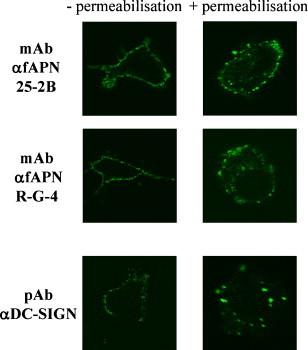
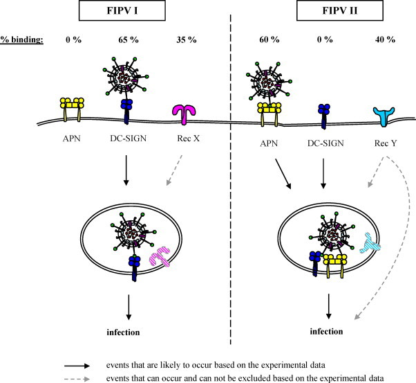
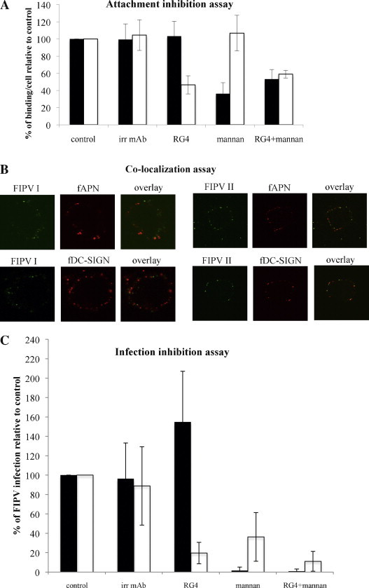
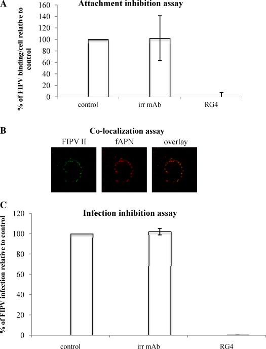
Two potential receptors have been described for the feline infectious peritonitis virus (FIPV): feline aminopeptidase N (fAPN) and feline dendritic cell-specific intercellular adhesion molecule grabbing non-integrin (fDC-SIGN). In cell lines, fAPN serves as a receptor for serotype II, but not for serotype I FIPV. The role of fAPN in infection of in vivo target cells, monocytes, is not yet confirmed. Both serotype I and II FIPVs use fDC-SIGN for infection of monocyte-derived cells but how is not known. In this study, the role of fAPN and fDC-SIGN was studied at different stages in FIPV infection of monocytes. First, the effects of blocking the potential receptor(s) were studied for the processes of attachment and infection. Secondly, the level of co-localization of FIPV and the receptors was determined. It was found that FIPV I binding and infection were not affected by blocking fAPN while blocking fDC-SIGN reduced FIPV I binding to 36% and practically completely inhibited infection. Accordingly, 66% of bound FIPV I particles co-localized with fDC-SIGN. Blocking fAPN reduced FIPV II binding by 53% and infection by 80%. Further, 60% of bound FIPV II co-localized with fAPN. fDC-SIGN was not involved in FIPV II binding but infection was reduced with 64% when fDC-SIGN was blocked. In conclusion, FIPV I infection of monocytes depends on fDC-SIGN. Most FIPV I particles already interact with fDC-SIGN at the plasma membrane. For FIPV II, both fAPN and fDC-SIGN are involved in infection with only fAPN playing a receptor role at the plasma membrane.
两种潜在的猫传染性腹膜炎病毒 (FIPV) 受体已被描述:猫氨基肽酶 N (fAPN) 和猫树突状细胞特异性细胞间黏附分子抓取非整联蛋白 (fDC-SIGN)。在细胞系中,fAPN 是血清型 II 的受体,但不是血清型 I FIPV 的受体。fAPN 在体内靶细胞单核细胞感染中的作用尚未得到证实。血清型 I 和 II FIPV 均使用 fDC-SIGN 感染单核细胞衍生细胞,但具体机制尚不清楚。在这项研究中,研究了 fAPN 和 fDC-SIGN 在 FIPV 感染单核细胞的不同阶段的作用。首先,研究了阻断潜在受体对附着和感染过程的影响。其次,确定了 FIPV 和受体的共定位水平。结果发现,阻断 fAPN 并不影响 FIPV I 的结合和感染,而阻断 fDC-SIGN 则将 FIPV I 的结合降低至 36%,并几乎完全抑制了感染。相应地,66%的结合 FIPV I 颗粒与 fDC-SIGN 共定位。阻断 fAPN 使 FIPV II 的结合减少 53%,感染减少 80%。此外,60%的结合 FIPV II 与 fAPN 共定位。fDC-SIGN 不参与 FIPV II 的结合,但阻断 fDC-SIGN 后感染减少了 64%。综上所述,FIPV I 感染单核细胞依赖于 fDC-SIGN。大多数 FIPV I 颗粒在质膜上已经与 fDC-SIGN 相互作用。对于 FIPV II,fAPN 和 fDC-SIGN 均参与感染,只有 fAPN 在质膜上发挥受体作用。