Systems Neurobiology Laboratories, Salk Institute for Biological Studies, 10010 North Torrey Pines Road, La Jolla, CA 92037, USA.
J Comp Neurol. 2012 Jan 1;520(1):52-80. doi: 10.1002/cne.22685.
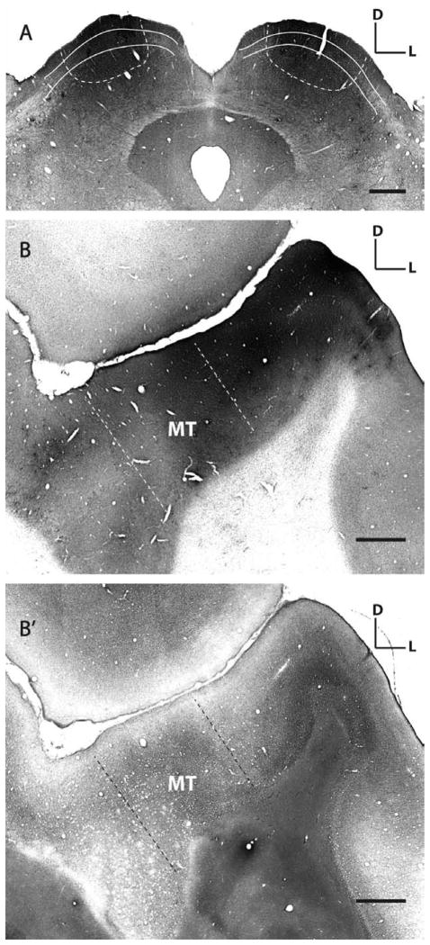
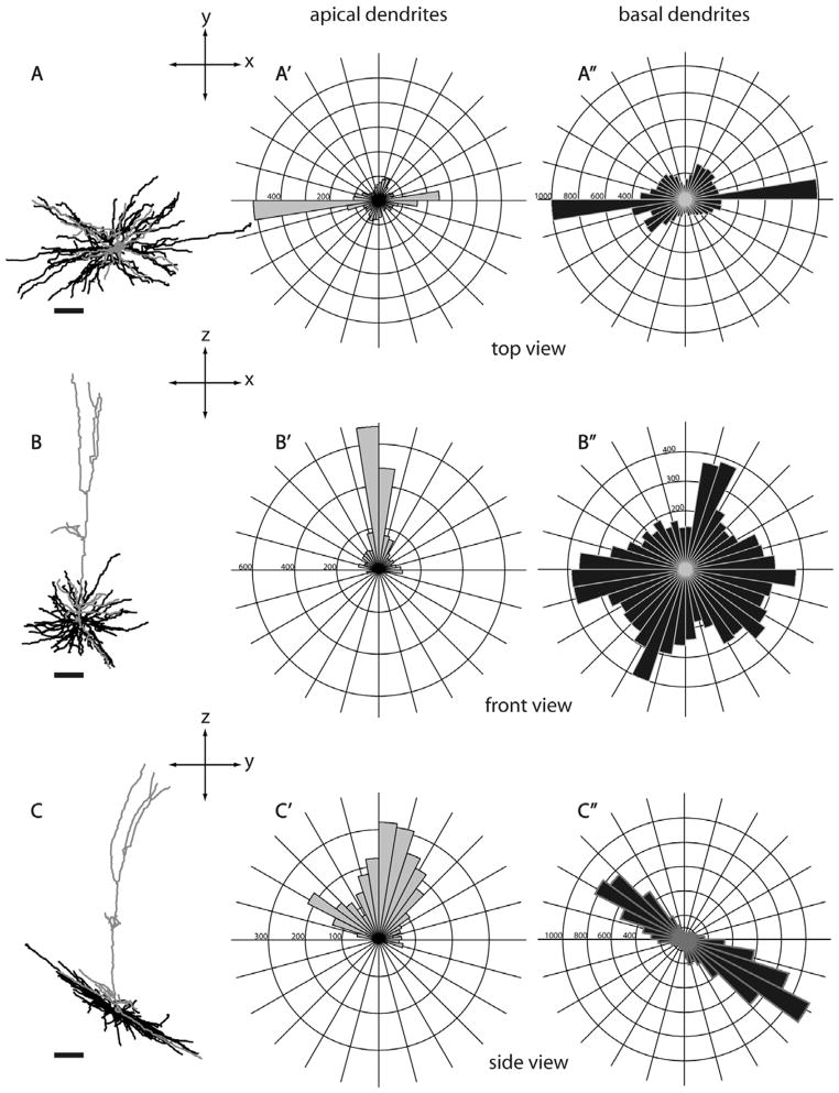
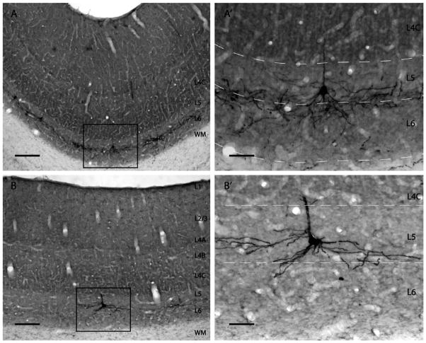
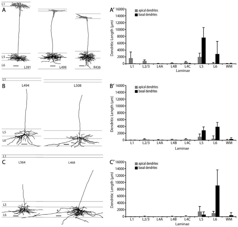
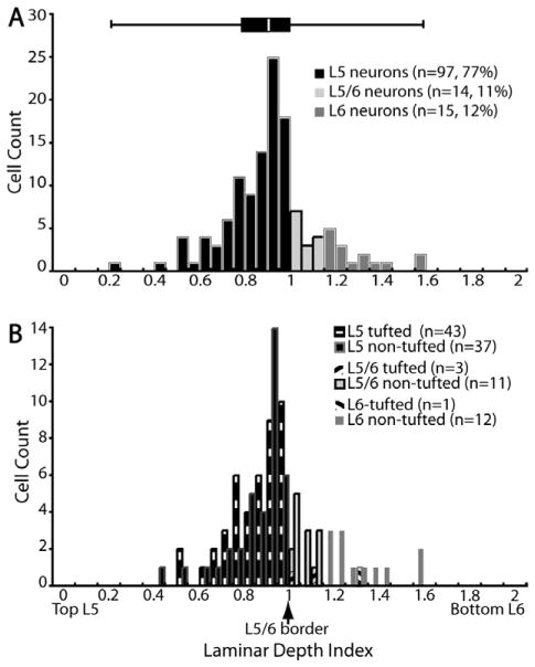
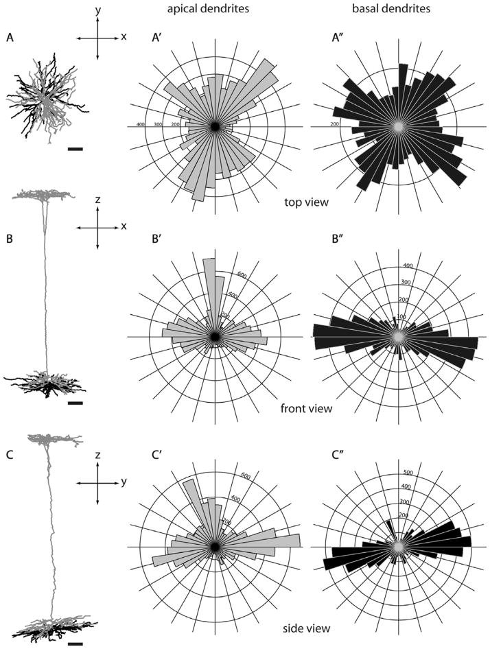
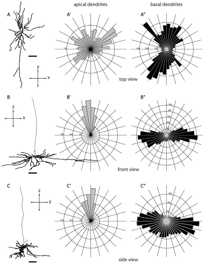
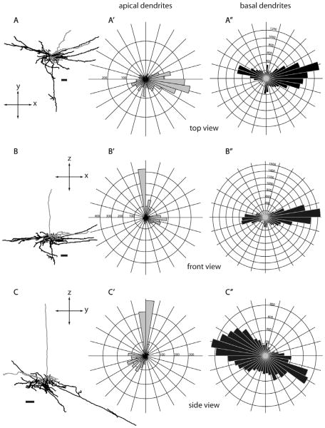
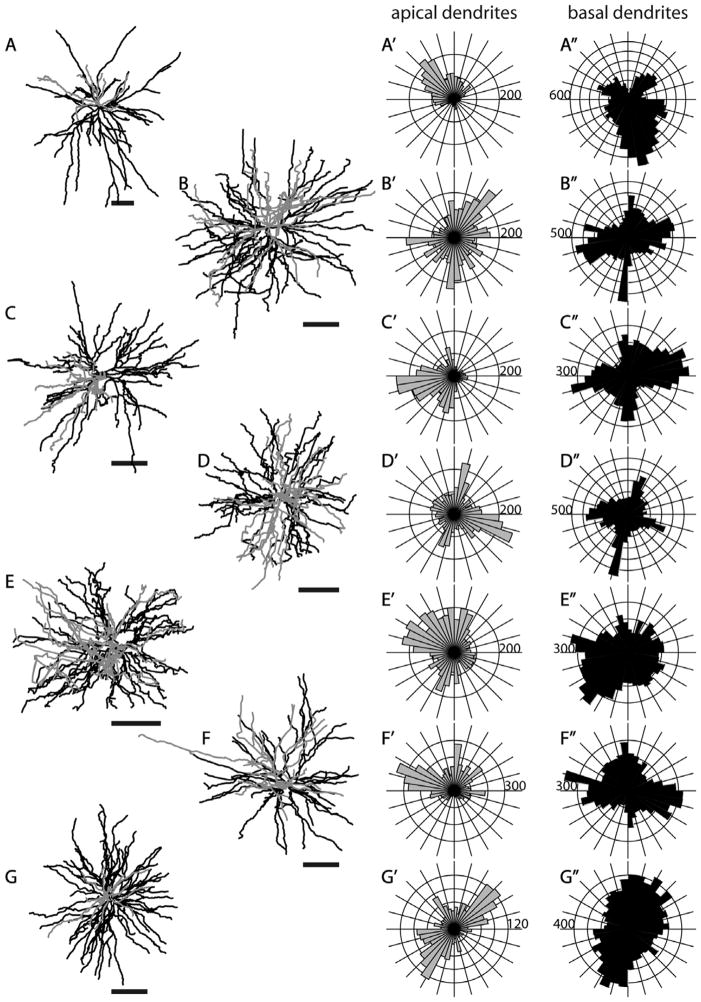
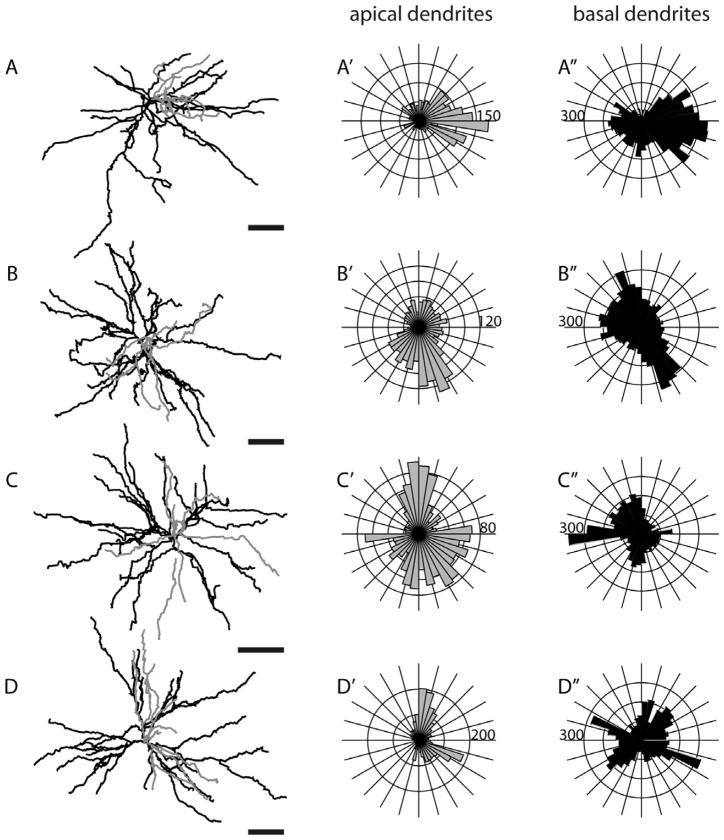
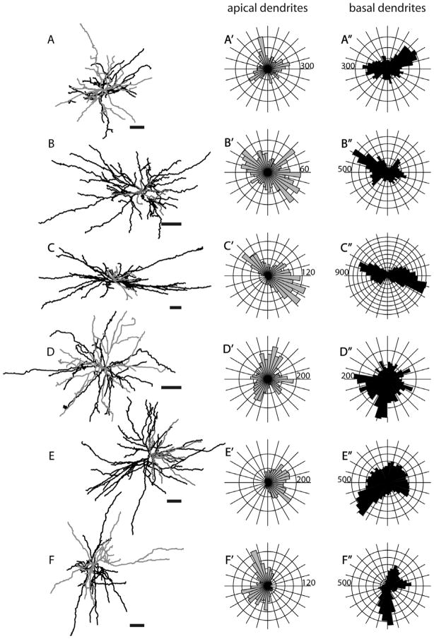
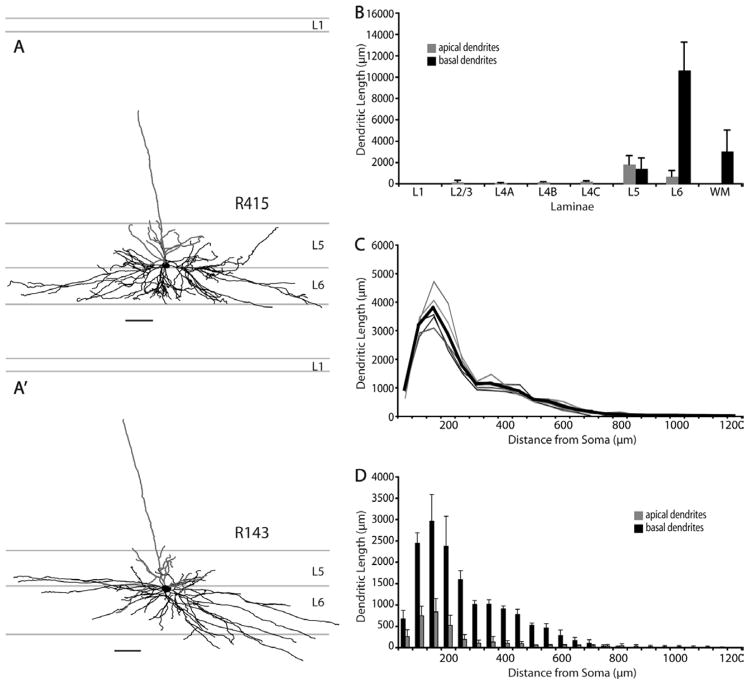
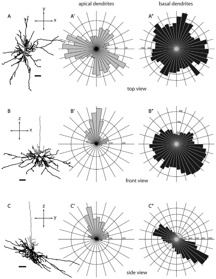
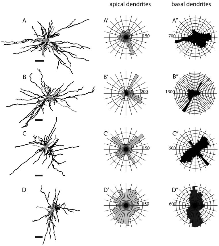
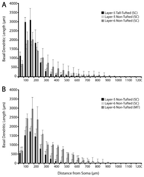
Layers 5 and 6 of primate primary visual cortex (V1) harbor morphologically diverse cell groups that have corticocortical and corticosubcortical projections. Layer 6 middle temporal area (MT)-projecting neurons are particularly interesting, as they are the only deep-layer cortical neurons that provide both corticocortical feedforward inputs (to area MT) and corticosubcortical feedback projections (to superior colliculus [SC]) (Fries et al. [1985] Exp Brain Res 58:613-616). However, due to limitations in anatomical tracing techniques, little is known about the detailed morphologies of these cells. We therefore applied a genetically modified rabies virus as a retrograde tracer to fill the dendrites of projection neurons with green fluorescent protein (GFP) (Wickersham et al. [2007] Nat Methods 4:47-49). We injected virus into SC or area MT of macaque monkeys and examined labeled cells in V1. Two-thirds of labeled neurons following SC injections were found in layer 5, consisting of "tall-tufted" and "nontufted" cells; the remaining cells were layer 6 "nontufted." Area MT injections labeled neurons in layers 4B and 6, as previously described (Shipp and Zeki [1989] Eur J Neurosci 1:309-332). The layer 6 neurons projecting to MT were remarkably similar to the layer 6 SC-projecting neurons. In contrast to the dense and focused dendritic arbors of layer 5 "tall-tufted" pyramids, all "nontufted" cells had sparse, but unusually long basal dendrites. The nontufted cells closely resemble Meynert cells (le Gros Clark [1942] J Anat 76:369-376; Winfield et al. [1983] Proc R Soc Lond B Biol Sci 217:129-139), but the full in vivo reconstructions presented here show that their basal dendrites can extend much further (up to 1.2 mm) and are less asymmetric than inferred from Golgi reconstructions.
灵长类动物初级视觉皮层(V1)的 5 层和 6 层拥有形态多样的细胞群,它们具有皮质间和皮质下投射。6 层中的颞中区域(MT)投射神经元特别有趣,因为它们是唯一的深层皮质神经元,它们提供皮质间的前馈输入(到区域 MT)和皮质下的反馈投射(到上丘 [SC])(Fries 等人,[1985]Exp Brain Res 58:613-616)。然而,由于解剖追踪技术的限制,对于这些细胞的详细形态知之甚少。因此,我们应用了一种经过基因改良的狂犬病毒作为逆行示踪剂,用绿色荧光蛋白(GFP)填充投射神经元的树突(Wickersham 等人,[2007]Nat Methods 4:47-49)。我们将病毒注射到猕猴的 SC 或区域 MT 中,并在 V1 中检查标记的细胞。SC 注射后的三分之二标记神经元位于 5 层,由“高丛状”和“非丛状”细胞组成;其余细胞为 6 层“非丛状”。如前所述,区域 MT 注射标记了 4B 层和 6 层的神经元(Shipp 和 Zeki,[1989]Eur J Neurosci 1:309-332)。投射到 MT 的 6 层神经元与投射到 SC 的 6 层神经元非常相似。与 5 层“高丛状”金字塔的密集和集中的树突相比,所有“非丛状”细胞都有稀疏但异常长的基底树突。非丛状细胞与 Meynert 细胞非常相似(le Gros Clark,[1942]J Anat 76:369-376;Winfield 等人,[1983]Proc R Soc Lond B Biol Sci 217:129-139),但这里呈现的完整体内重建显示,它们的基底树突可以延伸得更远(长达 1.2 毫米),并且不对称性比从高尔基重建推断的要小。