• 文献检索
  • 文档翻译
  • 深度研究
  • 学术资讯
  • Suppr Zotero 插件Zotero 插件
  • 邀请有礼
  • 套餐&价格
  • 历史记录
应用&插件
Suppr Zotero 插件Zotero 插件浏览器插件Mac 客户端Windows 客户端微信小程序
定价
高级版会员购买积分包购买API积分包
服务
文献检索文档翻译深度研究API 文档MCP 服务
关于我们
关于 Suppr公司介绍联系我们用户协议隐私条款
关注我们

Suppr 超能文献

核心技术专利:CN118964589B侵权必究
粤ICP备2023148730 号-1Suppr @ 2026

文献检索

告别复杂PubMed语法,用中文像聊天一样搜索,搜遍4000万医学文献。AI智能推荐,让科研检索更轻松。

立即免费搜索

文件翻译

保留排版,准确专业,支持PDF/Word/PPT等文件格式,支持 12+语言互译。

免费翻译文档

深度研究

AI帮你快速写综述,25分钟生成高质量综述,智能提取关键信息,辅助科研写作。

立即免费体验

核质 LIMK1 增强人乳腺癌的进展。

Nuclear and cytoplasmic LIMK1 enhances human breast cancer progression.

机构信息

Molecular Biology Program, University of Colorado Denver, 12801 East 17th Ave, Aurora, CO 80045, USA.

出版信息

Mol Cancer. 2011 Jun 18;10:75. doi: 10.1186/1476-4598-10-75.

DOI:10.1186/1476-4598-10-75
PMID:21682918
原文链接:https://pmc.ncbi.nlm.nih.gov/articles/PMC3131252/
Abstract

BACKGROUND

LIM kinase 1 (LIMK1) is expressed in both cytoplasmic and nuclear compartments, and is a key regulator of cytoskeletal organization involved in cell migration and proliferation. LIMK1 levels are increased in several human cancers, with LIMK1 over-expression in prostate and breast cancer cells leading to tumor progression. While it has been presumed that the mechanism by which LIMK1 promotes cancer progression is via its cytoplasmic effects, the role of nuclear vs cytoplasmic LIMK1 in the tumorigenic process has not been examined.

RESULTS

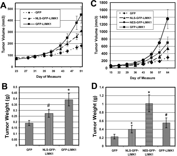
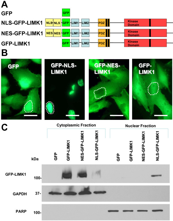
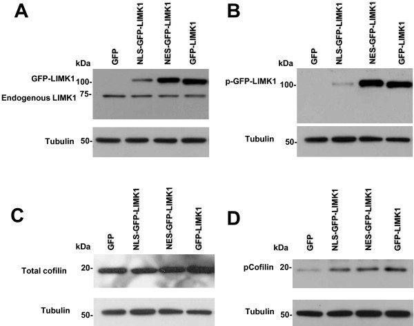
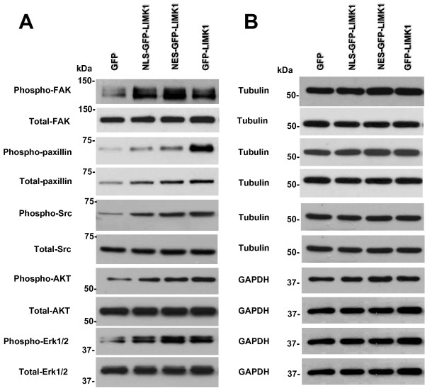
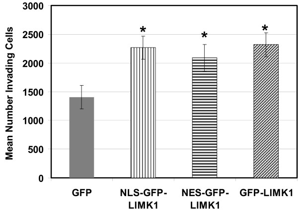
To determine if cytoplasmic or nuclear LIMK1 expression correlated with breast cancer, we performed immunohistochemical (IHC) analysis of breast tissue microarrays (TMAs), The IHC analysis of breast TMAs revealed that 76% of malignant breast tissue samples strongly expressed LIMK1 in the cytoplasm, with 52% of these specimens also expressing nuclear LIMK1. Only 48% of benign breast samples displayed strong cytoplasmic LIMK1 expression and 27% of these expressed nuclear LIMK1. To investigate the respective roles of cytoplasmic and nuclear LIMK1 in breast cancer progression, we targeted GFP-LIMK1 to cytoplasmic and nuclear subcellular compartments by fusing nuclear export signals (NESs) or nuclear localization sequences (NLS), respectively, to the amino-terminus of GFP-LIMK1. Stable pools of MDA-MB-231 cells were generated by retroviral transduction, and fluorescence microscopy revealed that GFP alone (control) and GFP-LIMK1 were each expressed in both the cytoplasm and nucleus of MDA-MB-231 cells, whereas NLS-GFP-LIMK1 was expressed in the nucleus and NES-GFP-LIMK1 was expressed in the cytoplasm. Western blot analyses revealed equal expression of GFP-LIMK1 and NES-GFP-LIMK1, with NLS-GFP-LIMK1 expression being less but equal to endogenous LIMK1. Also, Western blotting revealed increased levels of phospho-cofilin, phospho-FAK, phospho-paxillin, phospho-Src, phospho-AKT, and phospho-Erk1/2 in cells expressing all GFP-LIMK1 fusions, compared to GFP alone. Invasion assays revealed that all GFP-LIMK1 fusions increased MDA-MB-231 cell invasion ~1.5-fold, compared to GFP-only control cells. Tumor xenograft studies in nude mice revealed that MDA-MB-231 cells stably expressing GFP-LIMK, NLS-GFP-LIMK1 and NES-GFP-LIMK1 enhanced tumor growth 2.5-, 1.6- and 4.7-fold, respectively, compared to GFP-alone.

CONCLUSION

Taken together, these data demonstrate that LIMK1 activity in both the cytoplasmic and nuclear compartments promotes breast cancer progression, underscoring that nuclear LIMK1 contributes to the transforming function of LIMK1.

摘要

背景

LIM 激酶 1(LIMK1)表达于细胞质和核区室,是细胞迁移和增殖中细胞骨架组织的关键调节因子。在几种人类癌症中 LIMK1 水平增加,前列腺癌和乳腺癌细胞中的 LIMK1 过表达导致肿瘤进展。虽然人们推测 LIMK1 促进癌症进展的机制是通过其细胞质效应,但核区室与细胞质区室中 LIMK1 在肿瘤发生过程中的作用尚未被研究。

结果

为了确定细胞质或核区室 LIMK1 的表达是否与乳腺癌相关,我们对乳腺癌组织微阵列(TMA)进行了免疫组织化学(IHC)分析。对乳腺癌 TMA 的 IHC 分析表明,76%的恶性乳腺癌组织样本在细胞质中强烈表达 LIMK1,其中 52%的标本还表达核区室 LIMK1。只有 48%的良性乳腺样本显示强烈的细胞质 LIMK1 表达,其中 27%的标本表达核区室 LIMK1。为了研究细胞质和核区室 LIMK1 在乳腺癌进展中的各自作用,我们通过将核输出信号(NES)或核定位序列(NLS)分别融合到 GFP-LIMK1 的氨基末端,将 GFP-LIMK1 靶向到细胞质和核区室亚细胞区室。通过逆转录病毒转导生成 MDA-MB-231 细胞的稳定池,荧光显微镜显示 GFP 本身(对照)和 GFP-LIMK1 均在 MDA-MB-231 细胞的细胞质和核区室中表达,而 NLS-GFP-LIMK1 则在核区室中表达,NES-GFP-LIMK1 则在细胞质中表达。Western blot 分析显示 GFP-LIMK1 和 NES-GFP-LIMK1 的表达相等,NLS-GFP-LIMK1 的表达较少但与内源性 LIMK1 相等。此外,Western blot 分析显示,与 GFP 对照细胞相比,表达所有 GFP-LIMK1 融合蛋白的细胞中磷酸化 cofilin、磷酸化 FAK、磷酸化 paxillin、磷酸化 Src、磷酸化 AKT 和磷酸化 Erk1/2 的水平增加。侵袭实验显示,与 GFP 对照细胞相比,所有 GFP-LIMK1 融合蛋白均使 MDA-MB-231 细胞侵袭增加约 1.5 倍。裸鼠肿瘤异种移植研究显示,稳定表达 GFP-LIMK、NLS-GFP-LIMK1 和 NES-GFP-LIMK1 的 MDA-MB-231 细胞的肿瘤生长分别增强 2.5 倍、1.6 倍和 4.7 倍,与 GFP 对照相比。

结论

综上所述,这些数据表明,细胞质和核区室中的 LIMK1 活性均促进乳腺癌进展,突出表明核区室 LIMK1 有助于 LIMK1 的转化功能。

https://cdn.ncbi.nlm.nih.gov/pmc/blobs/1e36/3131252/bbdd501adfbb/1476-4598-10-75-7.jpg
https://cdn.ncbi.nlm.nih.gov/pmc/blobs/1e36/3131252/cbd736e8d72b/1476-4598-10-75-1.jpg
https://cdn.ncbi.nlm.nih.gov/pmc/blobs/1e36/3131252/f70b1a191e40/1476-4598-10-75-2.jpg
https://cdn.ncbi.nlm.nih.gov/pmc/blobs/1e36/3131252/d876d885fb1f/1476-4598-10-75-3.jpg
https://cdn.ncbi.nlm.nih.gov/pmc/blobs/1e36/3131252/f9eb114df5b7/1476-4598-10-75-4.jpg
https://cdn.ncbi.nlm.nih.gov/pmc/blobs/1e36/3131252/9d1cfa0d6059/1476-4598-10-75-5.jpg
https://cdn.ncbi.nlm.nih.gov/pmc/blobs/1e36/3131252/0ffff179b8b6/1476-4598-10-75-6.jpg
https://cdn.ncbi.nlm.nih.gov/pmc/blobs/1e36/3131252/bbdd501adfbb/1476-4598-10-75-7.jpg

相似文献

1
Nuclear and cytoplasmic LIMK1 enhances human breast cancer progression.核质 LIMK1 增强人乳腺癌的进展。
Mol Cancer. 2011 Jun 18;10:75. doi: 10.1186/1476-4598-10-75.
2
LIMK1 promotes the development of cervical cancer by up-regulating the ROS/Src-FAK/cofilin signaling pathway.LIMK1 通过上调 ROS/Src-FAK/cofilin 信号通路促进宫颈癌的发展。
Aging (Albany NY). 2024 Jul 5;16(13):11090-11102. doi: 10.18632/aging.206007.
3
LIM kinase 1 interacts with myosin-9 and alpha-actinin-4 and promotes colorectal cancer progression.LIM激酶1与肌球蛋白-9和α-辅肌动蛋白-4相互作用,并促进结直肠癌进展。
Br J Cancer. 2017 Aug 8;117(4):563-571. doi: 10.1038/bjc.2017.193. Epub 2017 Jun 29.
4
LIMK1 depletion enhances fasudil-dependent inhibition of urethral fibroblast proliferation and migration.LIMK1 耗竭增强了 fasudil 依赖型对尿道成纤维细胞增殖和迁移的抑制作用。
J Cell Biochem. 2019 Aug;120(8):12977-12988. doi: 10.1002/jcb.28569. Epub 2019 Mar 12.
5
Effect of manipulation on cartilage in rats with knee osteoarthritis based on the Rho-associated protein kinase/LIM kinase 1/Cofilin signaling pathways.基于 Rho 相关蛋白激酶/LIM 激酶 1/丝切蛋白信号通路探讨手法对膝骨关节炎大鼠软骨的影响。
J Tradit Chin Med. 2022 Apr;42(2):194-199. doi: 10.19852/j.cnki.jtcm.20220311.005.
6
LYRIC/AEG-1 is targeted to different subcellular compartments by ubiquitinylation and intrinsic nuclear localization signals.LYRIC/AEG-1 通过泛素化和内在核定位信号定位于不同的亚细胞区室。
Clin Cancer Res. 2009 May 1;15(9):3003-13. doi: 10.1158/1078-0432.CCR-08-2046. Epub 2009 Apr 21.
7
LIM kinase 1 increases tumor metastasis of human breast cancer cells via regulation of the urokinase-type plasminogen activator system.LIM激酶1通过调节尿激酶型纤溶酶原激活系统增加人乳腺癌细胞的肿瘤转移。
Int J Cancer. 2006 Jun 1;118(11):2703-10. doi: 10.1002/ijc.21650.
8
Effects of PAK4/LIMK1/Cofilin-1 signaling pathway on proliferation, invasion, and migration of human osteosarcoma cells.PAK4/LIMK1/Cofilin-1 信号通路对人骨肉瘤细胞增殖、侵袭和迁移的影响。
J Clin Lab Anal. 2020 Sep;34(9):e23362. doi: 10.1002/jcla.23362. Epub 2020 May 28.
9
Mapping of ESE-1 subdomains required to initiate mammary epithelial cell transformation via a cytoplasmic mechanism.通过细胞质机制启动乳腺上皮细胞转化所需的 ESE-1 亚结构域的作图。
Mol Cancer. 2011 Aug 28;10:103. doi: 10.1186/1476-4598-10-103.
10
Exosomal lncRNA H19 promotes the progression of hepatocellular carcinoma treated with Propofol via miR-520a-3p/LIMK1 axis.外泌体长链非编码 RNA H19 通过 miR-520a-3p/LIMK1 轴促进丙泊酚治疗的肝细胞癌的进展。
Cancer Med. 2020 Oct;9(19):7218-7230. doi: 10.1002/cam4.3313. Epub 2020 Aug 7.

引用本文的文献

1
Tetrahydropyrazolopyridinones as a Novel Class of Potent and Highly Selective LIMK Inhibitors.四氢吡唑并吡啶酮作为一类新型强效且高度选择性的LIMK抑制剂
J Med Chem. 2025 Aug 28;68(16):17427-17456. doi: 10.1021/acs.jmedchem.5c00974. Epub 2025 Aug 6.
2
New mechanistic understanding of FXR agonist Vonafexor: inducing sublethal damage of HBV-positive liver cancer cells via promoting anti-tumor immunity.法尼醇X受体激动剂沃纳法索对机制的新认识:通过促进抗肿瘤免疫诱导HBV阳性肝癌细胞的亚致死损伤。
Br J Cancer. 2025 Jun 30. doi: 10.1038/s41416-025-03089-z.
3
Identification of LIMK1 as a biomarker in clear cell renal cell carcinoma: from data mining to validation.

本文引用的文献

1
p57: A multifunctional protein in cancer (Review).p57:癌症中的多功能蛋白(综述)。
Int J Oncol. 2010 Jun;36(6):1321-9. doi: 10.3892/ijo_00000617.
2
Coagulation Factor Xa inhibits cancer cell migration via LIMK1-mediated cofilin inactivation.凝血因子 Xa 通过 LIMK1 介导的肌动蛋白丝切割蛋白失活抑制癌细胞迁移。
Thromb Res. 2010 Jun;125(6):e323-8. doi: 10.1016/j.thromres.2010.02.018. Epub 2010 Mar 26.
3
LIM-kinase is critical for the mesenchymal-to-amoeboid cell morphological transition in 3D matrices.LIM 激酶对于 3D 基质中的间质到阿米巴样细胞形态转变至关重要。
鉴定LIMK1作为透明细胞肾细胞癌的生物标志物:从数据挖掘到验证。
J Cancer Res Clin Oncol. 2025 Mar 8;151(3):104. doi: 10.1007/s00432-025-06146-5.
4
The dysadherin/FAK axis promotes individual cell migration in colon cancer.黏着斑失巢蛋白/粘着斑激酶轴促进结肠癌中单个细胞的迁移。
Int J Biol Sci. 2024 Apr 8;20(7):2356-2369. doi: 10.7150/ijbs.86699. eCollection 2024.
5
PDZ and LIM Domain-Encoding Genes: Their Role in Cancer Development.PDZ和LIM结构域编码基因:它们在癌症发展中的作用。
Cancers (Basel). 2023 Oct 19;15(20):5042. doi: 10.3390/cancers15205042.
6
Novel Histopathological Biomarkers in Prostate Cancer: Implications and Perspectives.前列腺癌中的新型组织病理学生物标志物:影响与展望。
Biomedicines. 2023 May 26;11(6):1552. doi: 10.3390/biomedicines11061552.
7
Retraction Note: α-santalol inhibits the angiogenesis and growth of human prostate tumor growth by targeting vascular endothelial growth factor receptor 2-mediated AKT/mTOR/P70S6K signaling pathway.撤稿说明:α-檀香醇通过靶向血管内皮生长因子受体2介导的AKT/mTOR/P70S6K信号通路抑制人前列腺肿瘤的血管生成和生长。
Mol Cancer. 2022 Dec 15;21(1):223. doi: 10.1186/s12943-022-01700-y.
8
Comparative Analysis of Small-Molecule LIMK1/2 Inhibitors: Chemical Synthesis, Biochemistry, and Cellular Activity.小分子 LIMK1/2 抑制剂的比较分析:化学合成、生物化学和细胞活性。
J Med Chem. 2022 Oct 27;65(20):13705-13713. doi: 10.1021/acs.jmedchem.2c00751. Epub 2022 Oct 7.
9
The microtubule cytoskeleton: An old validated target for novel therapeutic drugs.微管细胞骨架:新型治疗药物的一个古老且经证实的靶点。
Front Pharmacol. 2022 Sep 15;13:969183. doi: 10.3389/fphar.2022.969183. eCollection 2022.
10
LIMK1: A promising prognostic and immune infiltration indicator in colorectal cancer.LIMK1:结直肠癌中一个有前景的预后和免疫浸润指标。
Oncol Lett. 2022 May 30;24(1):234. doi: 10.3892/ol.2022.13354. eCollection 2022 Jul.
Biochem Biophys Res Commun. 2010 Feb 19;392(4):577-81. doi: 10.1016/j.bbrc.2010.01.075. Epub 2010 Jan 25.
4
A quantitative proteomics analysis of subcellular proteome localization and changes induced by DNA damage.定量蛋白质组学分析 DNA 损伤诱导的亚细胞蛋白质组定位和变化。
Mol Cell Proteomics. 2010 Mar;9(3):457-70. doi: 10.1074/mcp.M900429-MCP200. Epub 2009 Dec 21.
5
The Cdk inhibitor p57(Kip2) controls LIM-kinase 1 activity and regulates actin cytoskeleton dynamics.细胞周期蛋白依赖性激酶抑制剂p57(Kip2)可控制LIM激酶1的活性并调节肌动蛋白细胞骨架动力学。
Oncogene. 2009 Nov 26;28(47):4175-88. doi: 10.1038/onc.2009.269. Epub 2009 Sep 7.
6
Myocardin-related transcription factors and SRF are required for cytoskeletal dynamics and experimental metastasis.细胞骨架动力学和实验性转移需要心肌相关转录因子和血清反应因子。
Nat Cell Biol. 2009 Mar;11(3):257-68. doi: 10.1038/ncb1833. Epub 2009 Feb 8.
7
A tense situation: forcing tumour progression.一种紧张的情况:促使肿瘤进展。
Nat Rev Cancer. 2009 Feb;9(2):108-22. doi: 10.1038/nrc2544.
8
Downregulation of LIM kinase 1 suppresses ocular inflammation and fibrosis.LIM激酶1的下调可抑制眼部炎症和纤维化。
Mol Vis. 2008;14:1951-9. Epub 2008 Oct 30.
9
Nischarin inhibits LIM kinase to regulate cofilin phosphorylation and cell invasion.Nischarin抑制LIM激酶以调节丝切蛋白磷酸化和细胞侵袭。
Mol Cell Biol. 2008 Jun;28(11):3742-56. doi: 10.1128/MCB.01832-07. Epub 2008 Mar 10.
10
Expression of LIM kinase 1 is associated with reversible G1/S phase arrest, chromosomal instability and prostate cancer.LIM激酶1的表达与可逆性G1/S期阻滞、染色体不稳定和前列腺癌相关。
Mol Cancer. 2007 Jun 8;6:40. doi: 10.1186/1476-4598-6-40.