Structural Genomics Consortium, University of Toronto, Toronto, Ontario, Canada.
PLoS One. 2011;6(6):e18919. doi: 10.1371/journal.pone.0018919. Epub 2011 Jun 20.
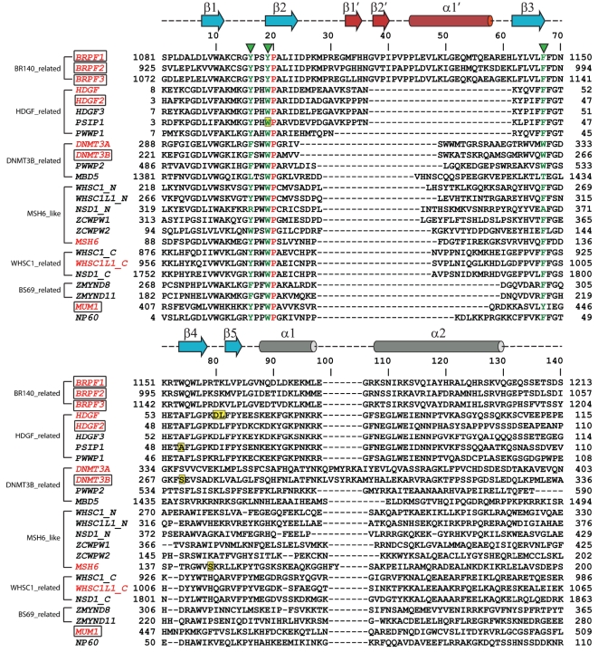
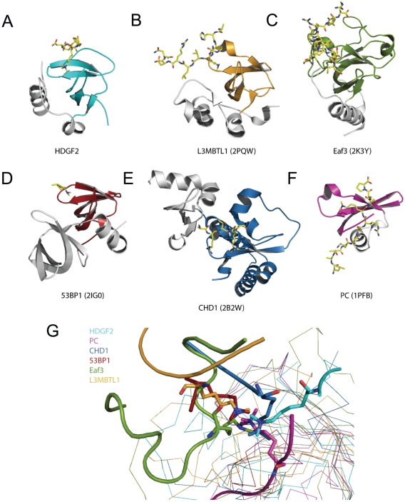
The PWWP domain was first identified as a structural motif of 100-130 amino acids in the WHSC1 protein and predicted to be a protein-protein interaction domain. It belongs to the Tudor domain 'Royal Family', which consists of Tudor, chromodomain, MBT and PWWP domains. While Tudor, chromodomain and MBT domains have long been known to bind methylated histones, PWWP was shown to exhibit histone binding ability only until recently.
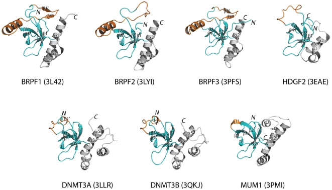
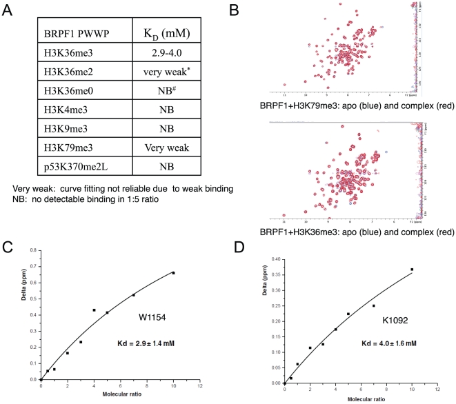
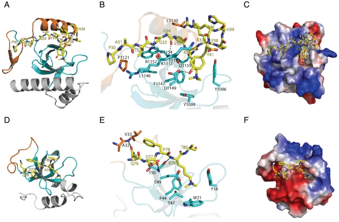
METHODOLOGY/PRINCIPAL FINDINGS: The PWWP domain has been shown to be a DNA binding domain, but sequence analysis and previous structural studies show that the PWWP domain exhibits significant similarity to other 'Royal Family' members, implying that the PWWP domain has the potential to bind histones. In order to further explore the function of the PWWP domain, we used the protein family approach to determine the crystal structures of the PWWP domains from seven different human proteins. Our fluorescence polarization binding studies show that PWWP domains have weak histone binding ability, which is also confirmed by our NMR titration experiments. Furthermore, we determined the crystal structures of the BRPF1 PWWP domain in complex with H3K36me3, and HDGF2 PWWP domain in complex with H3K79me3 and H4K20me3.
PWWP proteins constitute a new family of methyl lysine histone binders. The PWWP domain consists of three motifs: a canonical β-barrel core, an insertion motif between the second and third β-strands and a C-terminal α-helix bundle. Both the canonical β-barrel core and the insertion motif are directly involved in histone binding. The PWWP domain has been previously shown to be a DNA binding domain. Therefore, the PWWP domain exhibits dual functions: binding both DNA and methyllysine histones.
This article can also be viewed as an enhanced version in which the text of the article is integrated with interactive 3D representations and animated transitions. Please note that a web plugin is required to access this enhanced functionality. Instructions for the installation and use of the web plugin are available in Text S1.
PWWP 结构域最初在 WHSC1 蛋白中被鉴定为一个含有 100-130 个氨基酸的结构基序,被预测为一个蛋白-蛋白相互作用结构域。它属于 Tudor 结构域“皇家家族”,其中包含 Tudor、chromodomain、MBT 和 PWWP 结构域。虽然 Tudor、chromodomain 和 MBT 结构域早已被证实能与甲基化组蛋白结合,但直到最近才发现 PWWP 结构域具有组蛋白结合能力。
方法/主要发现:PWWP 结构域已被证明是一个 DNA 结合结构域,但序列分析和先前的结构研究表明,PWWP 结构域与其他“皇家家族”成员具有显著的相似性,这表明 PWWP 结构域有可能与组蛋白结合。为了进一步探索 PWWP 结构域的功能,我们使用蛋白质家族方法确定了来自七种不同人类蛋白的 PWWP 结构域的晶体结构。我们的荧光偏振结合研究表明,PWWP 结构域具有较弱的组蛋白结合能力,这也得到了我们的 NMR 滴定实验的证实。此外,我们还确定了 BRPF1 PWWP 结构域与 H3K36me3 的复合物以及 HDGF2 PWWP 结构域与 H3K79me3 和 H4K20me3 的复合物的晶体结构。
PWWP 蛋白构成了一个新的甲基赖氨酸组蛋白结合蛋白家族。PWWP 结构域由三个基序组成:一个典型的β-桶核心、第二个和第三个β-折叠之间的插入基序和一个 C 末端α-螺旋束。典型的β-桶核心和插入基序都直接参与组蛋白结合。PWWP 结构域先前被证明是一个 DNA 结合结构域。因此,PWWP 结构域具有双重功能:既结合 DNA,又结合甲基化赖氨酸组蛋白。